Radiology Reports
Radiology Report Format & Templates
A clear radiology report format helps radiologists present examination details, findings and impressions consistently. Explore downloadable radiology report samples for common X-ray and ult
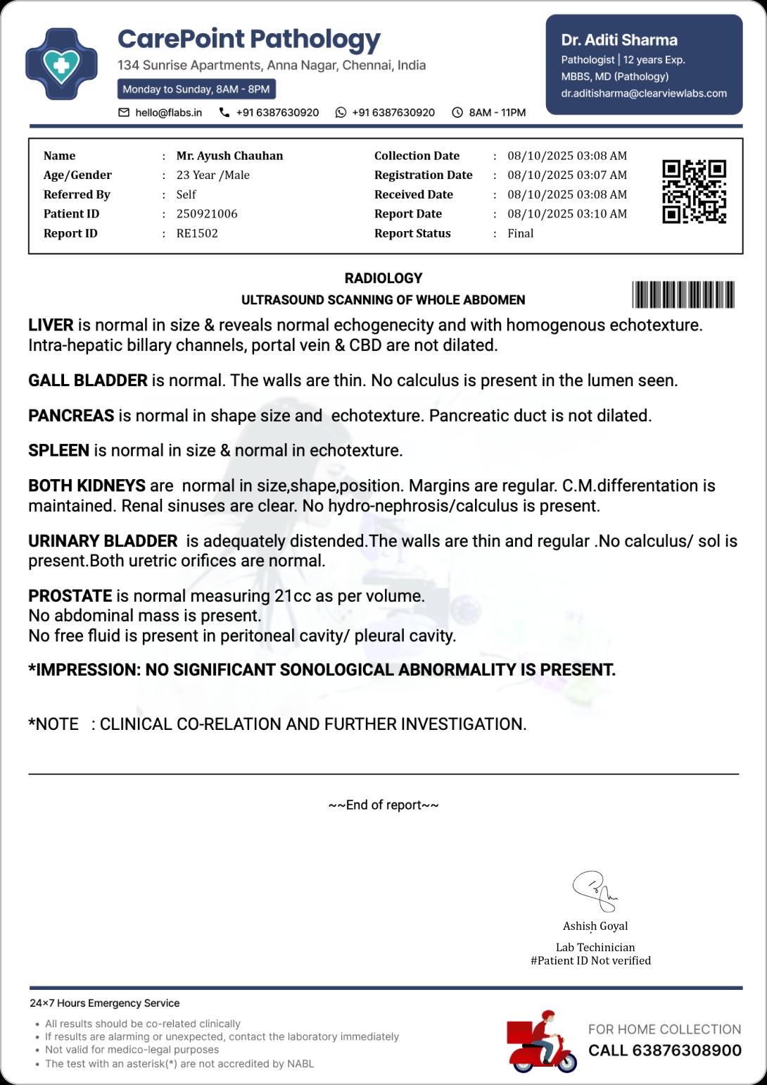
What Is Radiology Reporting?
Radiology reporting is the process of documenting the interpretation of diagnostic images such as X-rays, CT scans, MRI scans and ultrasounds.
Clinical indication
Why the scan was requested.
Examination technique
How the imaging was captured.
Imaging findings
What the images reveal.
Final impression
The summary for the referring doctor.
Download Radiology Report Formats and Templates
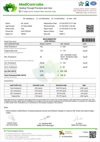
Browse downloadable radiology report formats for commonly performed X-ray and ultrasound examinations. Each sample can be used as a reference and should be adapted according to the examination and reporting protocol.
| S. No | Test Name | Download Sample Report |
|---|---|---|
| 1. | X RAY CHEST PA VIEW | Download PDF |
| 2. | X RAY CHEST AP VIEW | Download PDF |
| 3. | X RAY COCCYX AP & LATERAL VIEWS | Download PDF |
| 4. | X RAY COCCYX LATERAL VIEW | Download PDF |
| 5. | X RAY DORSAL SPINE AP AND LAT VIEW | Download PDF |
| 6. | X RAY DORSAL SPINE AP VIEW | Download PDF |
| 7. | X RAY ELBOW AP AND LAT VIEW | Download PDF |
| 8. | X RAY ELBOW AP VIEW | Download PDF |
| 9. | X RAY FISTULOGRAM | Download PDF |
| 10. | X RAY FOOT AP & LAT | Download PDF |
| 11. | X RAY FORE ARM AP AND LAT VIEW | Download PDF |
| 12. | X RAY HAND AP ANS OBLIQUE VIEW | Download PDF |
| 13. | X RAY HIP AP AND LATERAL VIEW | Download PDF |
| 14. | X RAY HSG | Download PDF |
| 15. | X RAY IVP | Download PDF |
| 16. | X RAY SKULL AP AND LAT VIEW | Download PDF |
| 17. | X RAY WRIST AP AND LAT VIEW | Download PDF |
| 18. | X RAY THIGH AP & LAT VIEW | Download PDF |
| 19. | X RAY PNS | Download PDF |
| 20. | X RAY PELVIS AP VIEW | Download PDF |
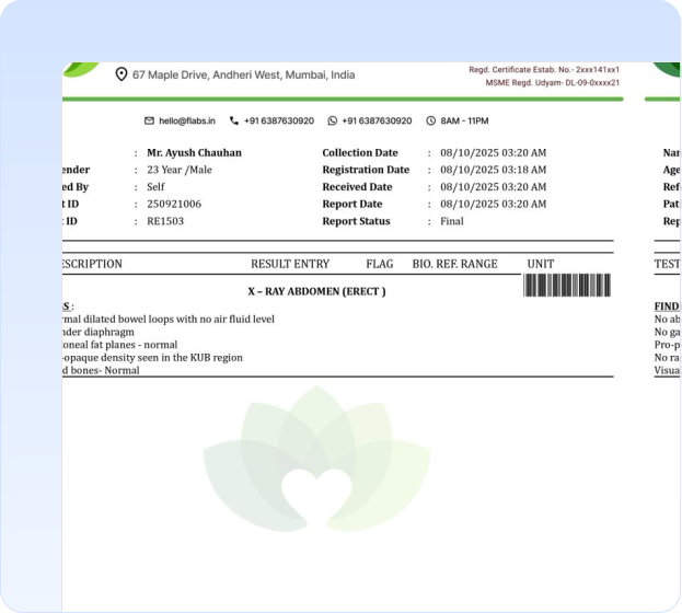
| 21. | X RAY ABDOMEN ( ERECT ) | Download PDF |
| 22. | X RAY FEET AP | Download PDF |
| 23. | ULTRASOUND PREGNANCY | Download PDF |
| 24. | ULTRASOUND ABDOMEN FEMALE (USF) | Download PDF |
| 25. | ULTRASOUND SCANNING OF EARLY TIFFA | Download PDF |
| 26. | ULTRASOUND BIOPHYSICAL PROFILE | Download PDF |
| 27. | ULTRASOUND CHEST & ABDOMEN | Download PDF |
| 28. | ULTRASOUND GUIDED FNAC | Download PDF |
| 29. | ULTRASOUND LEFT BREAST | Download PDF |
| 30. | ULTRASOUND SCAN OF EARLY PREGNANCY FINDINGS | Download PDF |
Sections in a Radiology Report Format
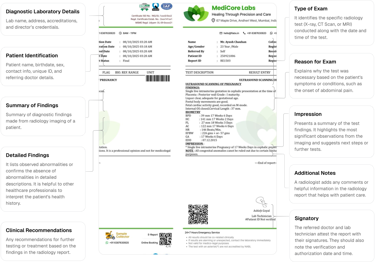
Diagnostic Laboratory Details
Lab name, address, accreditations, and director’s credentials.
Patient Identification
Patient name, birthdate, sex, contact info, unique ID, and referring doctor details.
Type of Exam
It identifies the specific radiology test (X-ray, CT Scan, or MRI) conducted along with the date and time of the test.
Reason for Exam
Explains why the test was necessary based on the patient's symptoms or conditions, such as the onset of abdominal pain.
Detailed Findings
It lists observed abnormalities or confirms the absence of abnormalities in detailed descriptions. It is helpful to other healthcare professionals to interpret the patient's health history.
Clinical Recommendations
Any recommendations for further testing or treatment based on the findings in the radiology report.
Summary of Findings
Summary of diagnostic findings made from radiology imaging of a patient.
Impression
Presents a summary of the test findings. It highlights the most significant observations from the imaging and suggests next steps or further tests.
Additional Notes
A radiologist adds any comments or helpful information in the radiology report that helps with patient care.
Signatory
The referred doctor and lab technician attest the report with their signatures. They should also note the verification and authorization date and time.
Why Use a Structured Radiology Report Format?
A structured radiology report format helps referring doctors locate the examination details, findings and impression quickly.
Standards in Radiology Reporting
Radiology reporting standards are crucial. They ensure consistency and precision in interpreting laboratory tests. They guarantee that radiologists deliver accurate and thorough information in their reports. This is key for diagnosis and effective treatment.
A well-designed radiology report example must stick to the standards below:
The UK's NICE (National Institute for Health and Care Excellence) releases global radiology reporting standards.
They emphasise structured lab reports, proper imaging use, and modality-specific guidelines. It also suggests that formats must show differences for different imaging techniques. For example, a CT report format is different from an X-ray report. The best radiology report formatting follows NICE's guidelines. But use them along with other standards and local protocols for best results.
Radiology Report Format: Best Practices
Standardised Structure
Ensure to use a standardised structure for all radiology reports in a consistent manner. It helps the involved healthcare professionals quickly find the information they need.
Clear and Concise Language
Avoid complex technical jargon that may confuse non-radiologist medical professionals. Use clear and concise language to describe findings, using globally accepted terminology by the concerned professionals.
Use Subheadings
Make the radiology report format organised with the use of subheadings to separate various sections. It enhances readability and allows the reader to easily navigate the report, especially in reports with multiple findings.
Use Multimedia
Try to incorporate images or hyperlinks to relevant images within the report. It greatly enhances the understanding and utility of the report, especially in digital radiology report formats generated through radiology report software
Patient Safety and Privacy
Ensure to maintain the patient privacy when designing a radiology report format. It must adhere to global and local medical regulations set by respective authorities (e.g ICMR and NABL in India).
Accessibility and Transferability
Well-designed radiology report formats should support transferability across healthcare bodies and technologies for seamless integration, ensuring easy access through different electronic health record (EHR) systems.
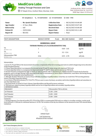
Mistakes to Avoid in Preparing Radiology Reports
- Lack of consistency in radiology report format layout and design.
- Not highlighting critical findings for improved visibility (e.g. bold fonts, italics, use of bullet points, etc.).
- Failure to update radiology report templates regularly.
- Overuse of Jargon and Acronyms.
- Excess metadata.
Radiology Report Format Standard in UK
1. Patient demographics
- Name
- Date of birth
- Sex
- Address
- Patient administration system (PAS)/ electronic patient record (EPR) number
- NHS number
2. Patient location at request
- Location description: ward name, etc
- Location type: A&E, inpatient, outpatient and GP
3. Requesting responsible consultant/GP
- ID-General Medical Council (GMC) number
- Name
- Job role as defined by the NHS Data Dictionary
- Main specialty as defined by the NHS Data Dictionary
- Employing institution as defined by the NHS Data Dictionary
4. Unique numbers
- Accession number = unique scheduling number issued by RIS
- Order number = Unique number issued by Ordercomms/electronic requesting system (RIS for paper requests)
5. Reporter (primary +/- secondary)
- ID-GMC number
- Name
- Job role of reporter as defined by the NHS Data Dictionary
- Main specialty as defined by the NHS Data Dictionary
- Employing institution as defined by the NHS Data Dictionary
6. Appointment date or study date (when exam was performed)
7. Exam room and institution
- Exam room and institution which owns the machine where the image acquisition took place (mobile scanners should be identified)
8. Date and time of primary report authorisation
9. Additional dates for corrections and report addenda
10. Priority
11. Patient category
12. Modality
13. Exam description
14. Where/to who copies of reports were sent
15. Report type
- Primary report
- Addendum
- Corrected primary report
- Corrected addendum
16. Fail-safe alert
- No alert
- Alert present
17. Narrative report text
Create Clear and Consistent Radiology Reports
A well-designed radiology report format helps diagnostic centres communicate findings clearly and maintain consistency across examinations.
Use the downloadable samples as references and adapt every radiology report template according to the examination and reporting practices of qualified professionals.
Frequently Asked Questions
A perfect radiology report example must include patient identity. It must also have health history, test results, and imaging techniques used. It must have detailed findings and lab information for reference.
DICOM stands for Digital Imaging and Communications in Medicine. It lays down a set of rules for exchanging decentralised data between healthcare professionals. A format for digital pictures guides how files organise medical images and related information.
ICMR (Indian Council of Medical Research) and NABL (National Accreditation Board for Testing and Calibration Laboratories) are the regulatory bodies in India that guide radiology reporting standards in India.
There are five main types of radiology: Diagnostic radiology, Interventional radiology, Nuclear medicine, Radiation therapy, and Ultrasound imaging.
X-ray, Ultrasound, CT scan, MRI, and PET scan are the most popular radiology lab tests. They diagnose various health conditions, such as cancer and haemorrhage.
A radiology report format is the structured layout used to document the clinical indication, technique, findings, impression and authorization details of an imaging examination.
A radiology report template is a reusable structure created for a particular imaging modality or examination.
It generally includes patient details, examination information, clinical indication, technique, findings, impression and radiologist details.
No. The format varies depending on the imaging modality, body part, clinical indication and reporting protocol.
Findings describe the imaging observations. The impression summarizes the most important diagnostic conclusions.
Yes. FLABS provides downloadable sample formats for several common X-ray and ultrasound examinations.
Yes. Imaging centres can customize the layout, centre information, examination fields, headings and signatory details.