
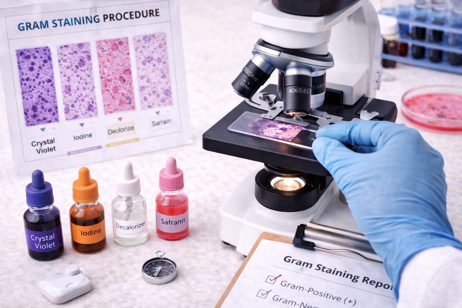
Laboratories rely on methods that allow fast differentiation of pathogens, and one technique continues to anchor diagnostic workflows: the Gram staining procedure. The method draws immediate attention because it delivers actionable insights within minutes.
It sparks interest during routine sample workups, holds attention through its structured sequence, and establishes confidence when Gram stain results align with clinical expectations. Pathologists and healthcare professionals rely on it every day to classify organisms, refine decisions, and support early treatment.
Origin of the Gram Staining Procedure
Hans Christian Gram introduced the Gram Staining method in 1884 while examining lung tissue from pneumonia patients. His objective was straightforward: create a staining method that improves the visualisation of bacteria within host tissue. The outcome became a foundational component of microbiology.
By design, the technique divides organisms based on cell wall structure. Gram-positive bacteria stain purple or blue due to their thick peptidoglycan layer.
Gram-negative stain reactions yield pink or red cells because their outer membrane loses the Crystal Violet–Iodine complex during alcohol exposure, allowing uptake of safranin.
Principle of Gram Staining in Microbiology
The Gram staining principle and procedure centre on the interaction between cell wall architecture and dye retention. Three factors influence this dynamic: lipid concentration in the outer membrane, peptidoglycan thickness, and permeability shifts on.
- Gram-positive organisms maintain a dense peptidoglycan network that traps the Crystal Violet–Iodine (CV-I) complex. Under oil immersion, they appear deep purple.
- Gram-negative organisms possess a thinner peptidoglycan layer and a high-lipid membrane. Alcohol dissolves this lipid barrier, allowing dye complexes to wash out. Once exposed to the safranin Gram stain, they turn pink.
Some laboratories also reference the pH theory of Gram staining, noting that cell wall charge dynamics influence affinity for cationic dyes. Although secondary to structural factors, it can explain occasional Gram-variable findings.
Gram Staining Requirements and Reagents
A consistent workflow depends on the correct set of Gram stain reagents. The Gram staining composition is uniform across facilities:
| Reagent | Role in the Gram Staining Procedure | Notes |
|---|---|---|
| Crystal Violet | Primary stain in the Gram stain | Stains all cells purple during the first step |
| Gram’s Iodine | Mordant | Forms large CV-I complexes that strengthen dye retention |
| Alcohol / Acetone | Decolourizer | Removes CV-I from Gram-negative organisms |
| Safranin | Counterstain | Colours decolourised cells pink or red |
Storage in dark bottles ensures dye stability. Fresh preparation avoids artefacts during Gram staining observation.
Slide Preparation
Accurate interpretation depends on smear quality. A thin, evenly spread suspension enhances clarity. Culture material mixes with a tiny drop of water, spreads gently, then air-dries. Heat fixing must be reserved and controlled because overheating distorts cells, influencing Gram stain interpretation.
Samples used for the Gram staining of bacteria experiment include sputum, throat swabs, urine, wound surfaces, blood cultures, or aspirates collected by fine-needle sampling. Each specimen must create a uniform film.
Gram Staining Process: Step-by-Step
The classical Gram staining technique relies on 4 sequential steps.
Primary stain: Flood the slide with Crystal Violet for thirty to sixty seconds, then rinse. All organisms appear purple at this stage.
Mordant application: Add Gram’s iodine for the same duration, then rinse. CV-I complexes strengthen within cell walls.
Decolourisation: Use alcohol or acetone. End the step when the runoff becomes clear. Timing varies by smear thickness and is the most sensitive phase in the entire Gram staining process. Over-decolourisation produces false Gram-negative outcomes, while under-decolourisation retains purple where it should not.
Counterstaining: Flood with safranin for thirty to sixty seconds, rinse gently, then dry.
Once complete,the examination shifts from 10X to 40X and finally to 100X oil immersion. Gram staining observation at high magnification reveals:
- Gram-positive cells: Purple
- Gram-negative cells: Pink or red
- Gram-staining fungi: Many yeasts appear Gram-positive due to thick walls.
What Happens in the Lab: Interpretation
Pathologists move beyond colour alone. Cell arrangement provides context. Cocci in chains suggest streptococci. Clusters point toward staphylococci. Rods with bipolar staining may raise suspicion for enteric pathogens.
The age of the culture influences outcomes. Older cultures behave irregularly and may produce Gram-variable organisms. Complex organisms such as Mycobacterium resist stains because of lipid-rich cell walls.
Environmental organisms like Acinetobacter may appear Gram-positive despite their Gram-negative nature. In uncertain cases, the KOH string test can support classification because Gram-negative bacteria lyse to produce a viscous thread.
Types of Gram Staining and Expanded Applications
Modified approaches extend the reach of the method. Heat-fixed tissue smears allow review of infected specimens. Automated staining systems standardise timing and volume for high-throughput labs. Cold-stain variations minimise reagent evaporation in warmer regions.
The broader application of Gram staining:
- Primary classification of bacterial pathogens.
- Early differentiation before culture growth becomes visible.
- Quality assessment of culture purity.
- Rapid orientation during sepsis workups.
- Initial screening of fungal elements in some cases
These outcomes support early decision-making in complex scenarios and complement advanced identification tools such as MALDI-TOF and molecular assays.
Gram Stain Results
Accurate identification of bacteria using staining techniques begins with dependable Gram stain results. The stain acts as the first branch in the decision tree that guides further biochemical assays, panels, and molecular interpretation.
Interpretation blends morphology, arrangement, staining intensity, and clinical context. Each smear becomes a preview of expected culture growth and antimicrobial susceptibility patterns. For laboratories managing high volumes, consistency has a direct influence on speed.
Strengthen Laboratory Operations With Flabs LIS
The full Gram staining procedure depends on standardised documentation, timed steps, and well-organised reporting structures. Modern laboratories benefit when these processes integrate smoothly into a centralised digital system.
Flabs Pathology Software, an AI-powered LIS platform, fits naturally within the Gram staining test by streamlining operational tasks from sample receipt to final reporting.
- Streamlined operations that align patient registration, automated analysis, and QR-coded report generation. There’s automated WhatsApp communication to reduce manual follow-ups.
- Enhanced patient experience through a dedicated portal where individuals view and download reports using their phone number login.
- A clean, intuitive interface that shortens onboarding time for technicians and pathologists.
- Scalable deployment that allows growth from single-site labs to multi-centre networks.
- Encrypted data management, controlled patient linkage, and routine backups.
Embedding Gram stain reporting templates inside the LIS can maintain structured Gram stain interpretation fields, reagent lot tracking, and timestamps. Laboratories that adopt Flabs notice smoother processes across operations.
Conclusion
The Gram staining procedure in microbiology connects cell wall architecture with rapid, interpretable visual cues. Hence, it is indispensable in microbiology labs. Each stage influences diagnosis. Pathologists rely on it to anchor decisions, preview organism behaviour, and shape various strategies.
Digital systems such as Flabs extend the technique’s value by providing structured workflows, faster reporting, and smoother communication. As microbiology continues to interface with automated platforms, Gram staining retains its relevance by adapting to both traditional bench techniques and modern LIS-driven environments.
Ready to Uplift Your Lab Procedures?
You can Start Your 5-Day Free Trial of Flabs Pathology Software by entering your information and exploring automated microbiology workflows firsthand. The platform maintains compliance standards and safeguards patient information at every step.
Get Started at ₹1!
Try Flabs for a full month for just ₹1.
Follow us on




