
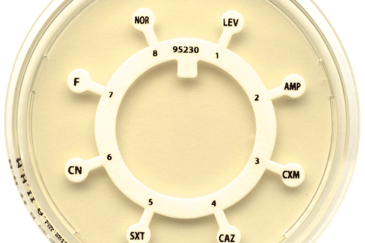
You receive a culture result in which the pathogen shows resistance to the first-line agent. You feel frustration as you look for actionable data in the report. You need clarity about which antimicrobial will work and which will not. You turn to the method lateral to dilutions—the one that, if done and interpreted properly, can steer therapy and laboratory decisions. The method is the “antibiotic disc” technique.
In your daily work as a pathologist or healthcare professional, you deal with the details of susceptibility testing. A solid grasp of the antibiotic disc process, including how discs are prepared, how to interpret results, and how to apply them in your setting, gives you confidence in your reports and consults. The goal here is to walk you through the method, interpretation and applications of the antibiotic disc approach in a clear, professional manner.
Note - The terms antibiotic disc, antibiotic discs, antibiotic disk, and antimicrobial disk are often used interchangeably in laboratory practice, although “disc” is the standard spelling in CLSI and EUCAST guidelines.
Antibiotic Disc: Method, Interpretation and Applications
An antibiotic disc is a small, sterile paper disc impregnated with a known concentration of an antimicrobial agent. When placed on agar inoculated with a bacterial isolate, the drug diffuses from the disc into the medium, setting up a concentration gradient. The area around the disc where bacterial growth is inhibited is called the zone of inhibition.
The method you employ involves these antibiotic discs for sensitivity testing. It is commonly called the disc diffusion method of antibiotic sensitivity testing. It allows you to categorise an isolate as susceptible, intermediate or resistant to specific antibiotics by measuring zone diameters and referencing standard breakpoints.
Antibiotic Disc Preparation and Setup
- Begin by selecting the appropriate medium. Most laboratories use Mueller‑Hinton agar (MHA) of standard thickness.
- Use Mueller-Hinton agar at pH 7.3 ± 0.1 and ensure the depth of the medium is about 4 mm. When testing Streptococcus or other fastidious species, supplement the medium with 5 % defibrinated sterile sheep blood before pouring plates.
- Prepare the inoculum in 5 mL of Trypticase Soy Broth and incubate 2–5 hours at 35–37 °C to achieve moderate turbidity.
- Ensure your bacterial suspension is adjusted to the 0.5 McFarland turbidity standard (equivalent to approx. 10⁸ CFU/mL) from a fresh culture.
- Once all discs are placed, allow a prediffusion time of 30 minutes at room temperature before incubation to ensure uniform diffusion.
- Swab the plate uniformly in two directions to create a confluent lawn. Allow the surface to dry for 3–5 minutes before placing discs.
- Place discs impregnated with known antibiotic concentrations (i.e., antibiotic discs for sensitivity testing) onto the agar surface with sterile forceps or applicator. Gently press to ensure full contact.
- Incubate plates at 35 ± 2 °C for 16–18 h (or per guideline) under aerobic conditions.
- After incubation measure the diameter of each inhibition zone (in mm) from edge to edge of clear zone including disc diameter. Use calipers/zone reader for accuracy.
Store antibiotic sensitivity discs at –20 °C to +8 °C and allow vials to reach room temperature before opening to prevent moisture condensation. After use, re-seal vials tightly and return them to refrigeration immediately.
For example, when you order an antibiotic disc for Gram-negative bacteria or an antibiotic disc for Gram-positive bacteria, you must ensure that the discs cover the spectrum of likely pathogens and resistance mechanisms in your institution.
Also, verify the size of the antibiotic disc (typical 6 mm diameter) and concentration (e.g., 10 µg, 30 µg depending on the antibiotic) matches guidelines.
Interpretation
Once zone diameters are measured, you compare the values against reference tables (from CLSI or EUCAST). The isolate is then reported as “susceptible”, “intermediate” or “resistant”.
In practice, when you use the antibiotic disc diffusion method, you interpret based on breakpoints. For example, if the zone diameter is equal to or greater than the susceptible cutoff, you report “S”; if it falls between susceptible and resistant cutoffs, then “I”; if below the resistant cutoff, you report “R”.
Remember that the zone diameter inversely correlates with the minimum inhibitory concentration (MIC) of the antibiotic for that isolate; larger zones generally indicate lower MICs, and vice versa.
Applications in Your Workflow
When you decide to order or interpret an antibiotic disc sensitivity test procedure, you apply it in the following settings.
Clinical Laboratories: You assess isolates from patient specimens to inform empiric and directed therapy.
Surveillance: You monitor resistance trends within Gram-negative and Gram-positive bacteria across time.
Research: You test new antimicrobial agents, combinations (though for combinations, other specialised methods may be needed).
When you select antibiotic discs names and panel compositions, tailor them to local formulary antimicrobials and prevalent pathogens. For example you may include cephalosporins, carbapenems, tetracyclines, etc., for Gram negative bacteria; and in Gram positives you include vancomycin, linezolid, daptomycin accordingly.
Another application: you might evaluate advanced resistance mechanisms like extended-spectrum β-lactamases (ESBL) or inducible clindamycin resistance using specialised disc diffusion configurations.
Nuances and Pitfalls
- Inoculum density too high or too low distorts zone sizes.
- Agar thickness or pH deviating from the standard alters diffusion and, therefore zone diameter.
- Incubation time and temperature must match guidelines to avoid false “susceptible”.
- Drug diffusion properties: Large or hydrophobic molecules diffuse slowly, yielding smaller zones independent of actual MIC.
Advantages and Limitations
| Advantages | Limitations |
|---|---|
| Simple and cost-effective method for assessing susceptibility across multiple isolates. | Provides categorical results only (no exact MIC values). |
| Widely standardised and reproducible under controlled laboratory conditions. | Sensitive to variations in inoculum, agar depth, and incubation environment. |
| Detects certain resistance mechanisms (e.g., ESBL, inducible clindamycin resistance). | Not suitable for fastidious or anaerobic organisms without modification. |
| Flexible panel design using antibiotic discs for Gram-positive and Gram-negative bacteria. | Slower turnaround compared to automated or rapid methods. |
| Supports surveillance and antimicrobial stewardship programs through trend monitoring. | Accuracy depends on disc potency, media quality, and adherence to procedure. |
Major Take-aways
- The antibiotic disc (paper disc impregnated with a known antibiotic), when used in the disc diffusion method, enables a robust way to classify isolates as susceptible, intermediate or resistant.
- Proper antibiotic disc preparation is foundational for accurate results.
- Interpretation hinges on measured zone diameters compared to standard breakpoints; you must integrate results with clinical and microbiological context.
- When you apply results, you think about both Gram-negative bacteria and Gram-positive bacteria panels, the panel of antibiotic discs names you include, and the surveillance implications for resistance.
- Be aware of variables that can invalidate results and collaborate with your laboratory to maintain high-quality protocols.
- As microbiology evolves, you remain apprised of emerging modifications (e.g., early growth disc diffusion) and continued standardisation efforts.
Common Antibiotic Discs for Sensitivity Testing
Antibiotic discs are used in laboratory sensitivity tests to determine how effective different antibiotics are against bacterial strains. Some commonly used discs include Amoxicillin, Ciprofloxacin, Gentamicin, Erythromycin, and Tetracycline. By placing these discs on bacterial cultures, scientists can observe zones of inhibition, which help identify the most effective antibiotic for treatment.
| Antibiotic Disc | Class | Common Use |
|---|---|---|
| Amoxicillin | Penicillin | Respiratory, Skin infections |
| Ciprofloxacin | Fluoroquinolone | Urinary tract infections, GI infections |
| Gentamicin | Aminoglycoside | Severe bacterial infections, Sepsis |
| Erythromycin | Macrolide | Respiratory infections, Skin infections |
| Tetracycline | Tetracycline | Acne, Respiratory infections |
In your role, you translate the data generated by the antibiotic disc diffusion method into actionable clinical consultations and reports. By understanding the method deeply, you strengthen your credibility and augment the utility of susceptibility data for clinicians, infection control, and microbiology teams.
Suggested - Petri Dish: Uses, Types, and Importance in Microbiology
Get Started at ₹1!
Try Flabs for a full month for just ₹1.
Follow us on




