
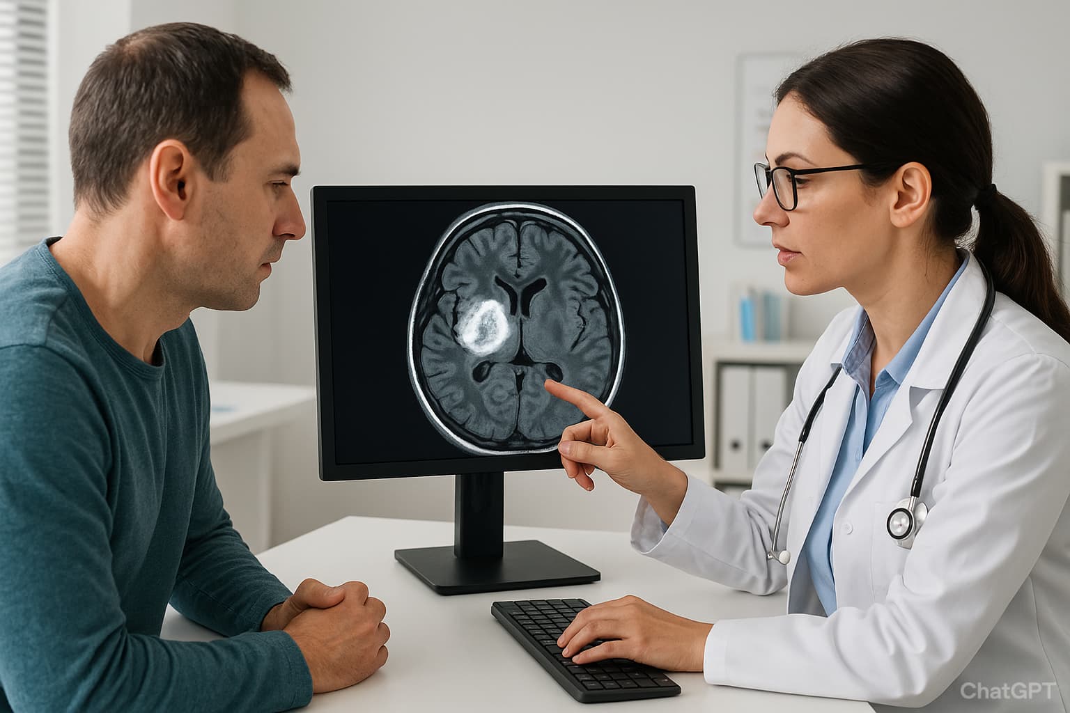
Brain tumours present a complex diagnostic challenge, requiring a combination of clinical evaluation, imaging, and laboratory techniques. For pathologists and healthcare professionals, a brain tumour test is not a single procedure but a structured set of diagnostic steps. These methods help establish the presence of abnormal tissue, determine tumour type, and guide therapeutic decisions.
This article explores the primary and advanced diagnostic pathways used in brain tumour assessment.
Brain Tumour Tests: Main Methods
| Test Name | Primary Role |
|---|---|
| Neurological Exam | Assesses reflexes, coordination, vision, and speech; helps identify localise dysfunction. |
| MRI (with contrast) | Gold standard; high-resolution imaging of brain tissue, detects tumour size, type, and spread. |
| Functional MRI (fMRI) | Maps blood flow during tasks to identify eloquent brain areas (speech, motor). |
| CT Scan | Provides quick brain images; detects bleeding, bone changes, or mass effect. |
| PET / PET-CT | Shows metabolic activity; differentiates tumour recurrence from treatment effect. |
| Stereotactic Biopsy | Confirms tumour type and grade through tissue sampling. |
| CSF Analysis / Lumbar Puncture | Detects malignant cells and biomarkers in cerebrospinal fluid. |
| Blood Tests & Molecular Markers | Supportive role; identify tumour-related hormones, genetic mutations, or biomarkers. |
Initial Clinical Assessment
The diagnostic pathway for a suspected brain tumour begins with a thorough clinical evaluation. Early bedside assessments can identify neurological deficits, localise the affected brain region, and guide the choice of subsequent imaging or laboratory tests.
Neurological Examination
The first step in any suspected case is a neurological exam, often considered the earliest brain tumour test in practice. Physicians assess:
- Reflexes (e.g., knee-jerk responses).
- Muscle strength and coordination.
- Balance and gait.
- Sensory responses to touch or pinpricks
- Cognitive performance (memory, arithmetic, problem-solving).
While a neurological exam cannot confirm a brain tumour, it helps localise possible areas of dysfunction (e.g., vision, hearing, balance, reflexes) and guides further testing.
An ophthalmoscopic exam is frequently included to check for optic nerve swelling, a sign of raised intracranial pressure.
Field Vision Test in Brain Tumour Cases
Visual pathway compromise is common in tumours affecting the occipital or pituitary regions. A field vision test for brain tumour detection helps evaluate blind spots and peripheral vision loss, which can be early indicators of mass effect.
Imaging-Based Brain Tumour Tests
MRI Scan
The most sensitive brain tumour test name clinicians rely on is MRI (Magnetic Resonance Imaging). MRI provides detailed anatomical images and is superior for differentiating tumour boundaries. It provides high-resolution brain parenchymal detail without ionising radiation. Gadolinium contrast improves lesion conspicuity and margin definition by highlighting areas of breakdown of the blood–brain barrier, clarifying lesion patterning that guides management.
Use of contrast agents highlights tumour vascularity and structural irregularities. There are some variants also.
MR Spectroscopy (MRS): Assesses chemical composition to differentiate tumour recurrence from necrosis.
MR Tractography: Maps neural tracts for surgical planning.
MR Perfusion Imaging: Evaluates tumour vascular supply.
In selected cases, a functional MRI (fMRI) may be performed to map regions responsible for speech, motor skills, or other critical functions before surgery. Incorporating fMRI into planning is associated with reduced perioperative adverse events, informing safer resection corridors.
Magnetic resonance perfusion can also quantify blood flow within different tumour regions, helping to distinguish aggressive from less active areas.
CT Scan
When an MRI is contraindicated (pacemakers, metal implants), a CT scan serves as an alternative. It provides rapid evaluation, e.g.,in cases of suspected haemorrhage or acute intracranial pressure rise.
PET and SPECT Scans
PET (Positron Emission Tomography): Highlights metabolic activity of tumour cells.
SPECT (Single Photon Emission CT): Assesses regional blood flow patterns.
PET scans are particularly useful for identifying fast-growing tumours such as glioblastomas or some oligodendrogliomas. However, slow-growing or benign tumours may not show up as clearly.
Laboratory and Blood-Based Brain Tumour Tests
While imaging remains central, interest in blood-based diagnostics is growing.
Brain Tumour Blood Test Possibilities
Most brain tumours cannot be definitively diagnosed with a simple blood test to detect a brain tumour, but certain markers and supportive investigations are useful.
Brain tumour marker test: Research is ongoing into identifying serum or CSF biomarkers such as GFAP (glial fibrillary acidic protein) and circulating tumour DNA.
Brain tumour genetic testing: Identifies mutations (e.g., IDH1, 1p/19q co-deletion, MGMT methylation) critical for prognosis and treatment decisions.
Brain tumour lab tests: Includes hormone assays in pituitary tumours or metabolic panels to evaluate systemic effects.
Brain tumour diagnosis blood tests: Can help exclude other causes of neurological symptoms or monitor systemic complications.
Tissue Sampling and Molecular Analysis
Tissue sampling methods in many ways are considered the decisive methods to identify brain tumours.
Biopsy
If imaging indicates a suspicious lesion, a biopsy remains the gold standard for confirming tumour histology. Pathologists examine tissue for malignancy, grade, and subtype. Biopsies may be obtained via Stereotactic needle procedures and Open surgical resections.
When open surgery is not feasible, a stereotactic needle biopsy may be performed. Contemporary series cite an approximate ~5% risk of bleeding complications with stereotactic needle biopsy.
Using CT or MRI guidance, a thin needle is inserted through a small opening in the skull to collect tissue. This approach reduces surgical risk, particularly for tumours in deep or critical brain areas.
Molecular and Genetic Testing
Modern brain tumour genetic testing allows clinicians to categorise tumours beyond histology. Identifying genetic signatures is essential in guiding targeted therapies and predicting prognosis.
Brain Tumour Grading
Tumours are graded from grade 1 (slow-growing, cells resembling normal tissue) to grade 4 (highly aggressive, poorly differentiated cells). Unlike many cancers, brain tumours are not staged, since they rarely spread outside the central nervous system. Grading provides essential prognostic information.
Cerebrospinal Fluid (CSF) Analysis
A lumbar puncture may be indicated in selected cases, especially for medulloblastoma or lymphoma. The CSF is examined for malignant cells and tumour markers. This method also assists in staging and monitoring relapse.
Brain Tumour Test Report and Interpretation
The brain tumour test report synthesises findings from multiple modalities:
- Imaging descriptions (size, location, vascularity).
- Histological classification.
- Molecular markers.
- Laboratory data (including relevant blood work).
Clinicians must correlate all results to produce a final diagnosis. Importantly, brain tumour test results are rarely standalone; they represent a diagnostic puzzle that integrates radiology, pathology, and laboratory sciences.
Brain Tumour Symptoms Test and Self-Assessment
Although patients often search for a brain tumour self-test or a brain tumour symptoms test, such approaches lack reliability. Symptoms such as persistent headaches, seizures, nausea, and vision disturbances should prompt immediate medical evaluation rather than self-assessment.
Healthcare professionals should caution patients against relying on online quizzes or self-screening tools.
Age and Screening in India
There is no universal age brain tumour screening test in India or globally. Screening is not recommended for asymptomatic individuals due to a lack of cost-effectiveness and low incidence. However, genetic counselling and testing may be indicated for families with inherited syndromes such as Li-Fraumeni or Turcot syndrome.
Brain Tumour and Blood Clots in the Brain Tests
Some tumours increase the risk of hypercoagulability, leading to venous sinus thrombosis. Diagnostic workups may therefore include:
- Coagulation profiles.
- MR venography or CT venography to detect clot formation.
Understanding the overlap is important, as clots can mimic tumour-related neurological symptoms.
Conclusion
A brain tumour test is not a singular diagnostic but a multidisciplinary process involving clinical, radiological, pathological, and molecular evaluations. From neurological exams and field vision testing to MRI, PET, and advanced brain tumour blood tests, each component builds towards a precise diagnosis.
For pathologists and healthcare professionals, integrating results into a comprehensive brain tumour test report ensures accurate classification and effective treatment planning. With growing interest in genetic profiling and biomarkers, the diagnostic landscape is evolving rapidly—making early, precise testing the cornerstone of improved patient outcomes.
Also check - Top 10 Radiology Software for Enhanced Diagnostic Imaging
Get Started at ₹1!
Try Flabs for a full month for just ₹1.
Follow us on




