
High-resolution computed Tomography (HRCT) of the thorax is a vital imaging technique for diagnosing and monitoring lung and chest diseases. Unlike conventional CT scans, HRCT provides detailed images with higher resolution, making it an essential tool for pathologists and healthcare professionals involved in respiratory diagnostics.
This article outlines the importance of the HRCT thorax test, its applications, procedure, interpretation and clinical relevance. We’ll also discuss the HRCT thorax test price, HRCT scan thorax usage, and the HRCT chest normal range to provide a complete understanding of the test.
What is an HRCT Thorax Test?
An HRCT thorax test is a specialised computed tomography (CT) scan that uses high-resolution imaging to assess lung structures in detail. This scan is commonly used for evaluating interstitial lung diseases (ILDs), infections, pulmonary fibrosis, and other respiratory conditions.
Unlike a standard CT scan, HRCT uses thin-section slices (typically 1–2 mm) and advanced image reconstruction techniques to enhance the visualisation of lung parenchyma, pleura, and airways.
HRCT Medical Abbreviation
HRCT stands for High-Resolution Computed Tomography. It is a specialized form of CT scan that provides highly detailed images of the lungs and chest, aiding in the accurate diagnosis of various respiratory conditions.
Why the HRCT Thorax Test is Done
Healthcare professionals recommend an HRCT scan thorax to detect and monitor conditions such as:
| Condition | Purpose of HRCT Thorax |
|---|---|
| Interstitial Lung Diseases (ILDs) | Identifies patterns of fibrosis, inflammation and scarring. |
| Pulmonary Fibrosis | Differentiates between idiopathic and secondary forms. |
| Chronic Obstructive Pulmonary Disease (COPD) | Assesses airway abnormalities. |
| Tuberculosis (TB) | Detects cavitations, nodules, and parenchymal damage. |
| Pneumonia and Lung Infections | Evaluate the extent and severity of infections. |
| Pulmonary Embolism | Assists in identifying vascular obstructions when combined with contrast-enhanced CT. |
| Lung Cancer Screening | Helps in detecting early-stage malignancies. |
| Occupational Lung Diseases | Used for evaluating asbestosis, silicosis, and hypersensitivity pneumonitis. |
| Emphysema | Detects alveolar damage and hyperinflation. |
| Bronchiectasis | Assesses airway dilation and mucus accumulation. |
| Hemoptysis | Evaluate the source of unexplained bleeding. |
HRCT Thorax Test Procedure
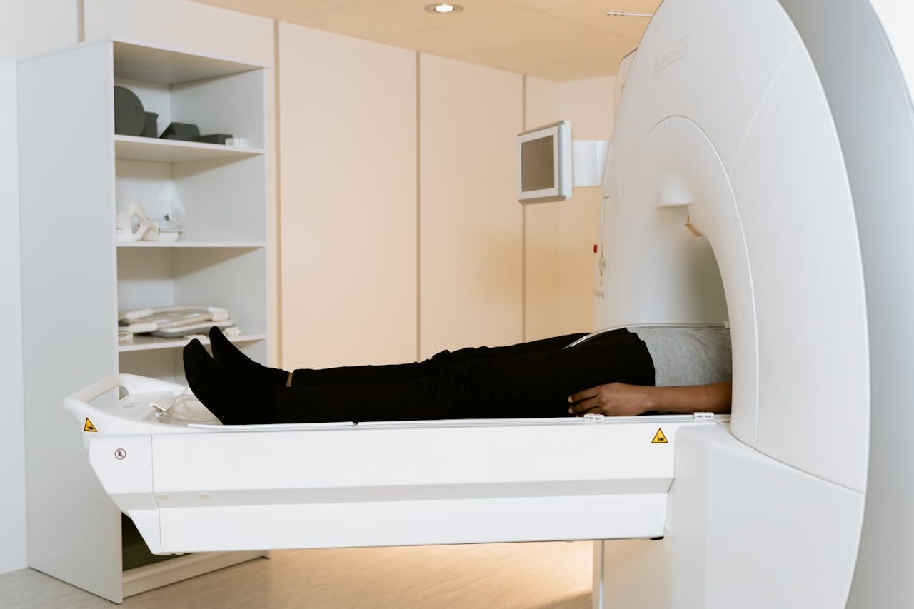
The HRCT scan thorax procedure is a quick and non-invasive imaging test. The patient lies flat on a motorised table that moves through the CT scanner. The scanner rotates around the chest, capturing multiple detailed X-ray images from different angles. These images are then processed by a computer to create high-resolution cross-sectional visuals of the thoracic region.
The scan itself is painless and takes just a few minutes. In some cases, a contrast dye may be injected into the patient’s vein to enhance the visibility of specific structures, such as blood vessels or abnormal growths. Patients are often instructed to hold their breath briefly during the scan to prevent motion artifacts and ensure clear imaging.
After the scan, patients can resume normal activities immediately unless contrast was used, in which case they may be monitored briefly for any allergic reactions.
The images are reviewed by a radiologist and sent to the referring physician for diagnosis and further evaluation.
When is HRCT Thorax Not Performed?
HRCT thorax is not performed if the patient is pregnant or suspected of pregnancy. Since the procedure involves radiation exposure, it is generally avoided unless absolutely necessary. Patients should also inform the healthcare provider about any allergies to contrast agents before undergoing a contrast-enhanced HRCT scan.
Interpretation
HRCT scans provide detailed images of the lung parenchyma, aiding in accurate diagnosis. The following findings help guide clinical decision-making:
1. HRCT Chest Normal Range
A normal HRCT chest scan should show clear lung fields, normal bronchovascular markings, and no abnormal nodules, fibrosis, or fluid accumulation. The pleura and mediastinum should appear normal without signs of thickening or effusion.
| Finding | Possible Indication |
|---|---|
| Ground-glass opacities | Early-stage infections, viral pneumonia, ILDs |
| Honeycombing | Pulmonary fibrosis, advanced ILDs |
| Nodules | Tuberculosis, lung cancer, granulomatous infections |
| Pleural thickening | Asbestosis, chronic inflammation |
| Air trapping | Obstructive lung disease (COPD, asthma) |
| Bronchiectasis | Recurrent infections, cystic fibrosis |
| Cavitary lesions | Tuberculosis, fungal infections, malignancy |
HRCT Thorax Test Price in India
The cost of an HRCT Thorax scan in India can range from ₹1,200 to ₹9,000, depending on the city, facility and whether contrast is used. In metro cities, the starting price is generally around ₹2,000 to ₹2,500, while in other regions, it may go up to ₹4,500 or more. Contrast-enhanced scans are usually priced higher, ranging from ₹4,500 to ₹9,000.
Prices can vary based on factors such as location, the quality of the facility, and additional services. Patients must ensure that they choose only the NABL-accredited medical laboratories for maximum safety.
Advantages of HRCT Thorax Over Standard CT
- Higher Resolution: Provides detailed lung imaging.
- Better Detection of ILDs: Essential for detecting subtle fibrosis patterns.
- Lower Radiation Dose: Uses thin-section imaging, reducing exposure compared to conventional CT.
- Ideal for Chronic Lung Disease Monitoring: Effective for tracking disease progression in conditions like pulmonary fibrosis and sarcoidosis.
Limitations of HRCT Thorax
- Limited Use for Soft Tissue Evaluation: Not ideal for mediastinal masses or vascular abnormalities.
- Potential Radiation Exposure: Though lower than standard CT, repeated scans should be justified.
- Contrast-Related Risks: Patients with kidney dysfunction may need alternative imaging options.
When Should HRCT Thorax Be Recommended?
A physician may recommend an HRCT scan thorax in cases of:
- Persistent respiratory symptoms (chronic cough, breathlessness, unexplained chest pain).
- Abnormal chest X-ray findings requiring further evaluation.
- Screening for occupational lung disease exposure.
- Follow-up for known lung disease progression or response to therapy.
Conclusion
For healthcare professionals, HRCT thorax remains a valuable tool for diagnosing and monitoring lung diseases. If you interpret HRCT scans, staying updated on abnormal patterns and disease markers enhances diagnostic accuracy. If you refer patients for HRCT, ensure they receive the right scan based on their symptoms and risk factors.
Discuss findings with radiologists when needed and use results to guide patient care effectively. Encourage them to take a second opinion if necessary. Keeping pace with advances in HRCT imaging will further improve diagnostic precision and treatment planning.
Also check - Understanding the Anatomy of a Normal Chest X-Ray
Get Started at ₹1!
Try Flabs for a full month for just ₹1.
Follow us on




