
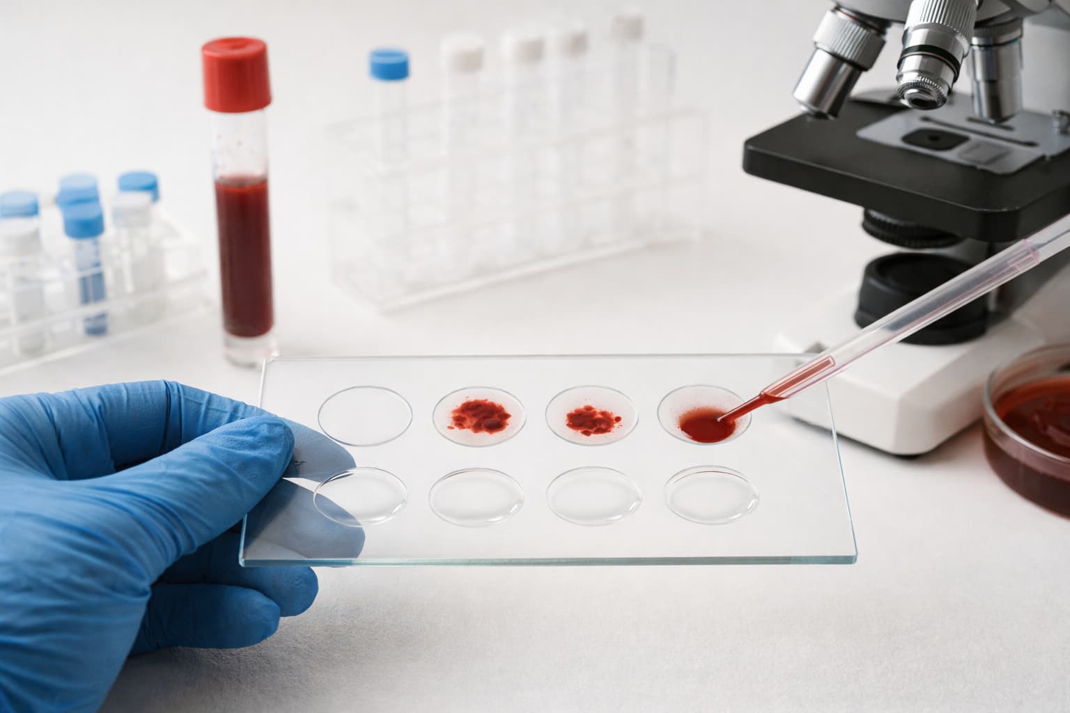
Agglutination testing keeps finding its way back into medical conversations because it remains surprisingly adaptable. Pathologists who want an assay that delivers clear, visual outcomes still lean on it for rapid clues about infectious diseases, immunology patterns, or blood group behaviors.
Many clinicians notice how it complements modern molecular diagnostics rather than competing with them. For labs balancing speed, cost, and dependable reaction visibility, the agglutination test is a familiar tool that deserves a fresh look.
Why the Standard Agglutination Test Still Matters
The agglutination test relies on a simple visual cue: clumping. When antibodies bind to particulate antigens , e.g., bacterial cells, latex beads, red blood cells, the reaction produces clusters that can be seen without sophisticated equipment.
The standard agglutination test (SAT) has remained in L1s, tertiary centers, and resource-limited settings because laboratories can deploy it with minimal infrastructure.
SAT is also a dependable serologic method for brucellosis, enteric fever panels, and a range of microbial evaluations. That familiarity builds confidence among healthcare teams who want assays with predictable performance.
Still, the SAT is only one piece of a larger ecosystem. Modern immunology uses variations each suited to a different purpose. Knowing how each variant behaves enables more precision in the procedure.
The Agglutination Principle: A Quick Refresher
The agglutination principle hinges on antigen–antibody interactions. When antibodies recognise surface antigens on cells or linked particles, cross-linking begins. Particles bridge together, forming clumps visible to the naked eye.
In agglutination immunology, those clumps indicate binding events that might otherwise require enzyme labels or fluorescent tags. The technique works especially well when dealing with particulate antigens. And for many microbial assays, that makes the agglutination reaction wonderfully direct.
The principle of agglutination reaction is influenced by antibody class and avidity, antigen density, environmental conditions such as pH or ion concentration, and reaction time.
Small shifts in any of these variables can alter the final outcome. When clumping fails to appear, analysts may mark it as “no agglutination,” meaning antibodies did not bind, antigen load was insufficient, or conditions were not ideal.
That absence can be just as informative as a strong positive.
Types of Agglutination Reactions
| Agglutination Type | How It Works | Notes |
|---|---|---|
| Direct Agglutination | Antibodies bind naturally occurring antigens on cells or particles. | Straightforward, relies on native surface antigens. |
| Passive Agglutination | Antigens are attached to carriers like latex beads. | Flexible format when natural particulate antigens are unavailable. |
| Reverse Passive Agglutination | Particles are coated with antibodies instead of antigens. | Useful when antigen detection is the priority. |
| Agglutination Inhibition Test | Soluble antigens in the sample block a visible reaction. | No clumping signals a positive result. |
| Slide Agglutination | Antigen–antibody reaction performed directly on a glass slide. | Favored for point-of-care or urgent bench work. |
| Tube Agglutination / Standard Agglutination Test (SAT) | Antigen–antibody reaction occurs in tubes with controlled volumes. | Provides semi-quantitative titers and structured interpretation. |
Agglutination Test in Microbiology
Microbiology labs use agglutination not for its immediate visual results. When organisms express surface antigens that respond predictably to targeted antibodies, agglutination reduces guesswork.
Salmonella and Brucella serology: SAT continues to serve as a dependable assay for quantification of antibody levels.
Neisseria and Streptococcus grouping: Slide techniques allow rapid classification steps during culture work-ups.
Latex-based passive agglutination for polysaccharide antigens such as pneumococcal or meningococcal markers.
These assays support faster decision-making in antimicrobial therapy selection and aid in the differentiation of visually similar organisms.
Precipitation vs Agglutination
Both rely on antigen–antibody interactions, yet they diverge in the type of antigen they detect.
| Feature | Precipitation | Agglutination |
|---|---|---|
| Antigen type | Soluble antigens | Particulate antigens |
| Visibility | Cloudiness or precipitate | Discrete clumps |
| Sensitivity | Lower | Higher |
| Common use | Immunodiffusion, immunoprecipitation | Microbial identification, serology |
| Equipment | Gel or liquid medium | Slides, tubes, or coated particles |
Many labs prefer agglutination techniques for fast interpretation when working with whole cells or synthetic carriers.
Medical Uses of the Agglutination Test
SAT has remained relevant in clinical practice due to its diagnostic versatility. While more advanced technologies have expanded the toolbox, SAT still holds its place in several key areas.
1. Brucellosis Serology
SAT quantifies antibodies against Brucella abortus or related species. Rising titers may suggest active infection, whereas declining titers might reflect recovery. In resource-limited regions, SAT sometimes becomes the mainstay diagnostic method.
2. Enteric Fever Evaluation
For laboratories using Widal-based testing panels, SAT is a structured method for assessing O and H antibody patterns against Salmonella enterica serovars. While molecular and culture methods bring higher specificity, SAT provides a quick serologic snapshot.
3. Blood Agglutination Test
Blood group determination relies on direct agglutination. Observing reactions between red cells and anti-A or anti-B reagents remains straightforward and widely used. The blood agglutination test also finds a role in assessing irregular antibodies in transfusion medicine.
Micro-opinions vary across institutions on ideal panel sizes.
4. Autoimmune and Immunologic Investigations
Agglutination methods can screen for rheumatoid factor, certain complement-related abnormalities, and antigen–antibody imbalances.
5. Rapid Pathogen Identification
Slide agglutination tests enable on-the-spot differentiation of bacterial isolates. Pathologists who want faster identification during outbreaks or time-sensitive cases often appreciate the familiarity of this method. It avoids delays associated with molecular workflows, especially during off-hours.
Interpreting Results
- Strong positive: Dense clumping forms quickly. Usually signals high antibody or antigen load.
- Weak positive: Fine granular clumps that might require careful tilting or gentle agitation.
- No agglutination, meaning there is either a lack of antibody–antigen interaction, inadequate antigen density, or an interfering factor.
False negatives arise from prozone effects, expired reagents, low-affinity antibodies, or inadequate incubation times. Technologists sometimes retest at different dilutions when results feel suspicious.
Final Thoughts
Agglutination assays and their variants continue to support medical diagnostics in ways that remain surprisingly relevant. For pathologists and healthcare professionals dealing with complex immunologic or microbiologic cases, the method still reveals clarity where it counts.
Knowing the nuances of antigen–antibody interactions, knowing how to interpret the clumping (or absence of it), and selecting the right agglutination format can improve diagnostic accuracy.
Also check - Treponemal Test: How It Detects Syphilis and What to Expect
Get Started at ₹1!
Try Flabs for a full month for just ₹1.
Follow us on




