
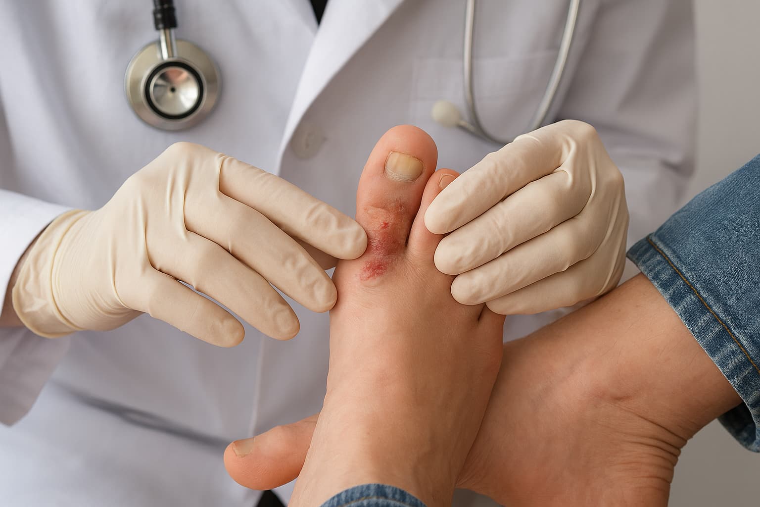
Superficial fungal infections can seem deceptively mild. Yet, beneath their visible manifestations lie subtle biological interactions between the human host and pathogenic fungi. These infections are limited to the outermost layers of skin, hair, and nails. Despite their limited depth, untreated cases may persist for years and influence diagnostic outcomes in dermatopathology and clinical microbiology.
Fungal organisms responsible for superficial infection thrive in warm and humid environments. In laboratories, specimens often show characteristic morphological patterns that distinguish one group from another.
Recognising those distinctions aids in diagnosis, guiding targeted antifungal management and reducing misclassification. Early recognition also prevents chronic colonisation and recurrent presentations in people.
Superficial Mycoses: Definition and Scope
Superficial mycoses refer to fungal infections confined to the stratum corneum, hair shaft, or nail surface without penetration into viable tissues. The infection remains non-inflammatory in most instances. From a microbiological viewpoint, these fungi utilise keratinised substrates as nutrient sources. But they rarely provoke host immune response due to the absence of tissue invasion.
Classification of Mycoses
| Category | Site of Infection | Example Organisms | Clinical Involvement |
|---|---|---|---|
| Superficial Mycoses | Stratum corneum, hair, nails | Malassezia, Trichosporon, Piedraia hortae, Hortaea werneckii | Cosmetic or asymptomatic |
| Cutaneous Mycoses | Epidermis, hair follicles, nails | Trichophyton, Microsporum, Epidermophyton | Inflammatory response, tissue damage |
| Subcutaneous Mycoses | Dermis, subcutaneous tissue | Sporothrix schenckii, Cladosporium carrionii | Chronic nodules or abscesses |
| Systemic Mycoses | Internal organs | Histoplasma capsulatum, Blastomyces dermatitidis | Disseminated infections |
Superficial infection remains confined to the first category. Its study is essential for differentiating non-invasive keratinophilic fungi from deeper, tissue-invasive pathogens.
Classification of Superficial Mycoses
The classification of superficial mycoses relies on site involvement and fungal morphology. Four principal superficial mycoses types are recognised.
- Pityriasis Versicolor (Tinea versicolor): Caused by Malassezia species. The yeast colonises sebaceous areas, producing hypopigmented or hyperpigmented macules with fine scaling. Lesions fluoresce under Wood’s lamp with a yellowish-green hue.
- Tinea Nigra: Caused by Hortaea werneckii. Presents as solitary brown to black macules on palms or soles. No scaling or inflammation is evident.
- Black Piedra: Caused by Piedraia hortae. Involves hard, black nodules attached to scalp hair shafts.
- White Piedra: Caused by Trichosporon species. Forms soft, white nodules along hair shafts of beard, moustache, or genital areas.
Each entity displays a unique morphology of superficial mycoses in laboratory examination. Direct microscopy of KOH-treated samples and culture on Sabouraud agar reveal the structural variations defining each pathogen.
Superficial Mycoses Causative Agents
The superficial mycoses causative agent varies by infection type.
- Malassezia furfur – lipid-dependent yeast producing “spaghetti and meatballs” appearance on microscopy.
- Hortaea werneckii – dematiaceous fungus forming brown septate hyphae and yeast-like cells.
- Piedraia hortae – pigmented hyphae forming hard, cemented nodules around hair.
- Trichosporon beigelii – yeast with hyaline hyphae and arthroconidia in white piedra.
The superficial mycoses microbiology reveals their adaptation to keratinised environments. Most species rely on lipids or keratin as substrates. They lack aggressive proteolytic enzymes, which limit tissue invasion. Growth requirements in vitro often include lipid supplementation for Malassezia species or specific pH adjustment for Hortaea werneckii.
Superficial Mycoses Symptoms
Superficial mycoses symptoms remain confined to cosmetic or surface alterations.
- Skin lesions: Discolouration, fine scaling, or macular patches without erythema.
- Hair involvement: Nodules, brittleness, or hair shaft breakage.
- Nail changes: Discolouration limited to the surface plate without onycholysis.
- No systemic signs: No fever, malaise, or lymphadenopathy.
While patients may remain asymptomatic, dermatopathologists must differentiate lesions from melanocytic or bacterial conditions during examination.
Diagnosis of Superficial Mycoses
Accurate identification requires correlation of clinical and laboratory findings. The laboratory diagnosis of superficial mycoses involves multiple steps.
Specimen Collection: Skin scrapings from active margins, affected hair, or nail fragments. Sterile instruments minimise contamination.
Direct Microscopy: Potassium hydroxide (10–20%) mount for keratin digestion. Observation of hyphae, budding yeast, or arthroconidia patterns confirms fungal presence.
Culture: Inoculation on Sabouraud dextrose agar, with or without lipid supplementation, assists in isolation. Colony morphology and pigmentation are diagnostic clues.
Microscopic Identification: Lactophenol cotton blue staining or Gram staining delineates hyphal septation and spore arrangement.
Malassezia shows clusters of round yeast with short hyphal fragments.
Piedraia shows pigmented filamentous forms.
Molecular Techniques: PCR and MALDI-TOF MS improve identification accuracy and species-level discrimination.
Morphology of Superficial Mycoses
Microscopically, fungi causing superficial infections demonstrate varied structural arrangements:
- Malassezia species: Short, curved hyphae with round budding yeast cells.
- Hortaea werneckii: Branched, septate hyphae with dark pigment granules.
- Trichosporon: Hyaline, septate hyphae producing arthroconidia.
- Piedraia hortae: Pigmented, tightly compacted hyphae forming cemented nodules.
These morphological patterns correspond to specific infection sites and influence diagnostic interpretation. The correlation between the morphology of superficial mycoses and clinical presentation remains essential for precise classification.
Pathogenesis and Transmission
Transmission occurs through direct contact, contaminated fomites, or self-inoculation. Humidity, sebum production, poor hygiene, and immunosuppression are some of the factors that promote colonisation. Pathogenic fungi adhere to keratinised surfaces via specific adhesins and produce enzymes that modify surface lipids. However, they rarely invade deeper layers due to host barriers and their limited enzymatic profile.
Treatment
Effective superficial mycoses treatment depends on the infection type and the extent of involvement. Therapy targets fungal eradication while preserving skin integrity.
Topical Therapy
First-line management involves topical antifungals.
- Azoles such as ketoconazole, clotrimazole, and econazole.
- Allylamines such as terbinafine and naftifine.
- Ciclopirox olamine or selenium sulfide for Malassezia infections.
Treatment duration ranges from one to four weeks. Cleansing with antifungal shampoos containing zinc pyrithione benefits scalp and trunk involvement.
Systemic Therapy
Oral agents may be considered for extensive or recurrent infections.
- Itraconazole or fluconazole for pityriasis versicolor unresponsive to topical therapy.
- No systemic treatment is necessary for tinea nigra or piedra unless refractory.
Adjunctive Measures
Hygiene optimisation, avoidance of occlusive clothing, and disinfection of personal items prevent reinfection. Hair trimming facilitates the removal of nodules in piedra.
Prevention and Implications
Preventive strategies revolve around limiting moisture accumulation, controlling sebum production, and maintaining skin hygiene. In hospital settings, differentiation of superficial infection from nosocomial fungal colonisation prevents misdirected antifungal use. Accurate laboratory reporting supports dermatologists and infectious disease specialists in targeted management.
For pathologists, awareness of superficial mycoses aids in tissue section interpretation. Occasionally, superficial colonisation may appear in biopsy specimens from unrelated dermatologic conditions. Recognising their non-invasive morphology prevents diagnostic confusion with cutaneous or subcutaneous mycoses.
Conclusion
Superficial mycoses remain confined to the most accessible human tissues yet possess distinctive microbiological signatures that require careful evaluation. Knowledge of their classification, morphology, and causative agents supports precise diagnosis and treatment.
For healthcare professionals, integrating clinical observation with laboratory data ensures accurate differentiation from other dermatomycoses. Effective management reduces recurrence and maintains diagnostic accuracy within pathology and clinical microbiology practice.
Also check - Glass Slide: Types and Role in Microscopy & Histology
Get Started at ₹1!
Try Flabs for a full month for just ₹1.
Follow us on




