
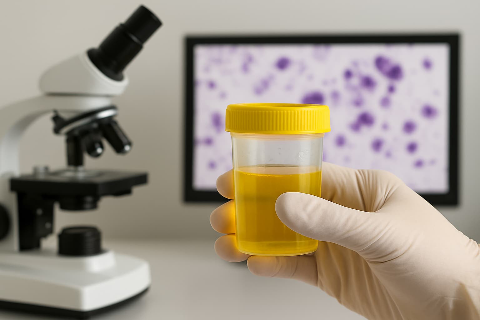
Hematuria and unexplained irritative voiding symptoms present a frequent diagnostic conundrum in urology and primary care. While the differential diagnosis ranges from benign infection to malignancy, failing to identify high-grade urothelial carcinoma early leads to poor prognostic outcomes.
Invasive procedures like cystoscopy are definitive. But they impose patient discomfort and utilise significant resources, making them less ideal as a sole primary screening tool. The urine cytology test serves as a potent, non-invasive adjunct. The test also has high specificity for high-grade lesions and enables pathologists to detect cellular abnormalities before they become visible on gross inspection.
Scope of Urine Cytology Test
A urine cytology test involves the microscopic evaluation of exfoliated cells found in voided urine or washing specimens. The primary objective differs from a standard urinalysis; while the latter looks for chemical properties and infection markers, cytology focuses specifically on cytomorphology to identify neoplastic cells originating from the urothelium lining the bladder, ureters, renal pelvis, and urethra.
For pathologists, the assay is particularly effective for detecting high-grade urothelial carcinoma (HGUC) and carcinoma in situ (CIS), which shed cells more readily than low-grade tumours. It acts as a complement to cystoscopy and imaging, filling diagnostic gaps where flat lesions (CIS) might be missed visually but detected via cellular shedding.
Specimen Collection and Pre-Analytical Variables
The accuracy of cytology test results depends heavily on specimen quality. Urothelial cells degenerate rapidly in urine due to low pH and bacterial proliferation.
The "First Morning" Myth
Contrary to many other urine tests, first-morning urine is unsuitable for cytology. Urine held in the bladder overnight causes significant cellular degradation, rendering the urine slide under the microscope unreadable or artefact-prone. The optimal specimen is a second voided midstream sample, collected after hydration to ensure fresh, well-preserved cells.
Collection Methods
1. Voided Urine: The most common non-invasive method. Requires a clean-catch technique to minimise contamination from squamous epithelial cells (skin/vaginal) or bacteria.
2. Catheterised Specimen: Used when patients cannot void voluntarily. Pathologists must be informed of this method, as instrumentation often causes the shedding of cell clusters (instrumentation artefact) that mimics cancer.
3. Bladder Washings: Saline is vigorously flushed into the bladder and retrieved. It yields a higher cellularity and better preservation than voided urine. But it is invasive.
Laboratory Processing
Upon receipt, the laboratory concentrates the sample. Modern pathology lab equipment, e.g., cytocentrifuges or liquid-based cytology processors (e.g., ThinPrep), deposits a monolayer of cells onto a glass slide. These are stained (usually with Papanicolaou stain) to highlight nuclear detail, chromatin patterns, and nuclear-to-cytoplasmic ratios.
The Paris System for Reporting Urinary Cytology
To standardise reporting and reduce ambiguity, the pathology community widely adopts The Paris System (TPS). The framework focuses on the detection of HGUC.
Interpretation of Results
| TPS Category | Cytomorphologic Findings | Clinical Significance |
|---|---|---|
| Negative for High-Grade Urothelial Carcinoma (NHGUC) | Benign urothelial cells, squamous cells, and inflammatory cells. | No evidence of high-grade malignancy. Does not rule out low-grade tumours. |
| Atypical Urothelial Cells (AUC) | Mild nuclear enlargement, hyperchromasia, or irregular borders. | "Grey zone." Cells are not normal but fall short of malignancy criteria. Requires close follow-up. |
| Suspicious for High-Grade Urothelial Carcinoma (SHGUC) | Some cells show severe atypia (high N/C ratio, coarse chromatin), but the quantity is low. | High probability of cancer. Warrant immediate cystoscopic evaluation. |
| High-Grade Urothelial Carcinoma (HGUC) | Distinctly malignant cells: enlarged nuclei, irregular membranes, coarse chromatin. | Diagnostic of cancer. Proceed to staging/treatment. |
| Unsatisfactory/ Non-Diagnostic | Low cellularity, obscuring blood/inflammation, or lubricant contamination. | The test must be repeated. |
Analysing Microscopic Findings
When evaluating a urine slide under a microscope, the cytopathologist looks for specific deviations from normal cellular architecture.
Epithelial Cells and Atypia
Finding epithelial cells positive in urine is normal; the urinary tract constantly sheds lining cells. The distinction lies in the morphology. Normal urothelial cells are small with smooth nuclear membranes. In contrast, malignant cells exhibit:
- Increased Nuclear-to-Cytoplasmic (N/C) ratio.
- Hyperchromasia (dark staining nuclei).
- Irregular nuclear borders.
Decoy Cells and Viral Mimics
A specific diagnostic pitfall involves decoy cells in urine. These are urothelial cells infected with Polyomavirus (BK virus), commonly seen in kidney transplant recipients or immunocompromised patients. They contain large, ground-glass viral inclusions that can mimic high-grade cancer cells. Distinguishing these requires a trained eye or adjunctive immunostaining (SV40 stain) to prevent a false-positive cancer diagnosis.
Limitations of the Test
The urine cytology test is not a perfect catch-all. Its performance is stratified by tumour grade.
High-Grade Tumours/CIS: Sensitivity is high (roughly 90-95%). These aggressive cells lack cohesion and shed easily into the urine, making cytology an excellent detection tool.
Low-Grade Tumours: Sensitivity is low (10-50%). Low-grade papillary urothelial neoplasms usually consist of cohesive cells that look nearly identical to normal urothelium. A negative cytology result does not rule out low-grade bladder cancer.
Therefore, a negative result in a patient with gross hematuria still necessitates cystoscopy to visualise potential low-grade papillary tumours.
Cost and Turnaround
For healthcare administrators and patients, logistical factors are relevant. The turnaround time for urine cytology is generally rapid— results are available within 24 to 48 hours, depending on the need for ancillary studies or second opinions.
The urine cytology test cost varies by institution, geography, and insurance coverage. It generally costs between ₹176 and ₹1,800 in India. The cost is significantly lower than molecular marker tests or invasive cystoscopy.
Follow-Up
Management relies on the integration of cytology test results with clinical findings.
- Negative Result: In high-risk patients (e.g., history of bladder cancer), routine surveillance continues. In symptomatic patients, imaging (CT Urogram) and cystoscopy are used to rule out low-grade lesions.
- Atypical (AUC): It can trigger a repeat test in 3 months or immediate reflex testing with fluorescence in situ hybridisation (FISH) to assess chromosomal abnormalities.
- Suspicious/Positive: Immediate cystoscopy is mandatory. If the bladder appears normal on white-light cystoscopy, physicians may employ blue-light cystoscopy or take random bladder biopsies to locate carcinoma in situ (CIS).
Conclusion
The urine cytology test provides a non-invasive window into the cellular dynamics of the urinary tract. Thus, it enables the early detection of high-grade malignancies that pose the greatest threat to patient survival. For a pathologist, strict adherence to specimen preparation protocols and the Paris System reporting standards ensures that this tool delivers maximum clinical utility, guiding urologists toward appropriate intervention and surveillance strategies.
Also check - Coagulogram Test: A Complete Guide to Clotting Tests
Get Started at ₹1!
Try Flabs for a full month for just ₹1.
Follow us on




