
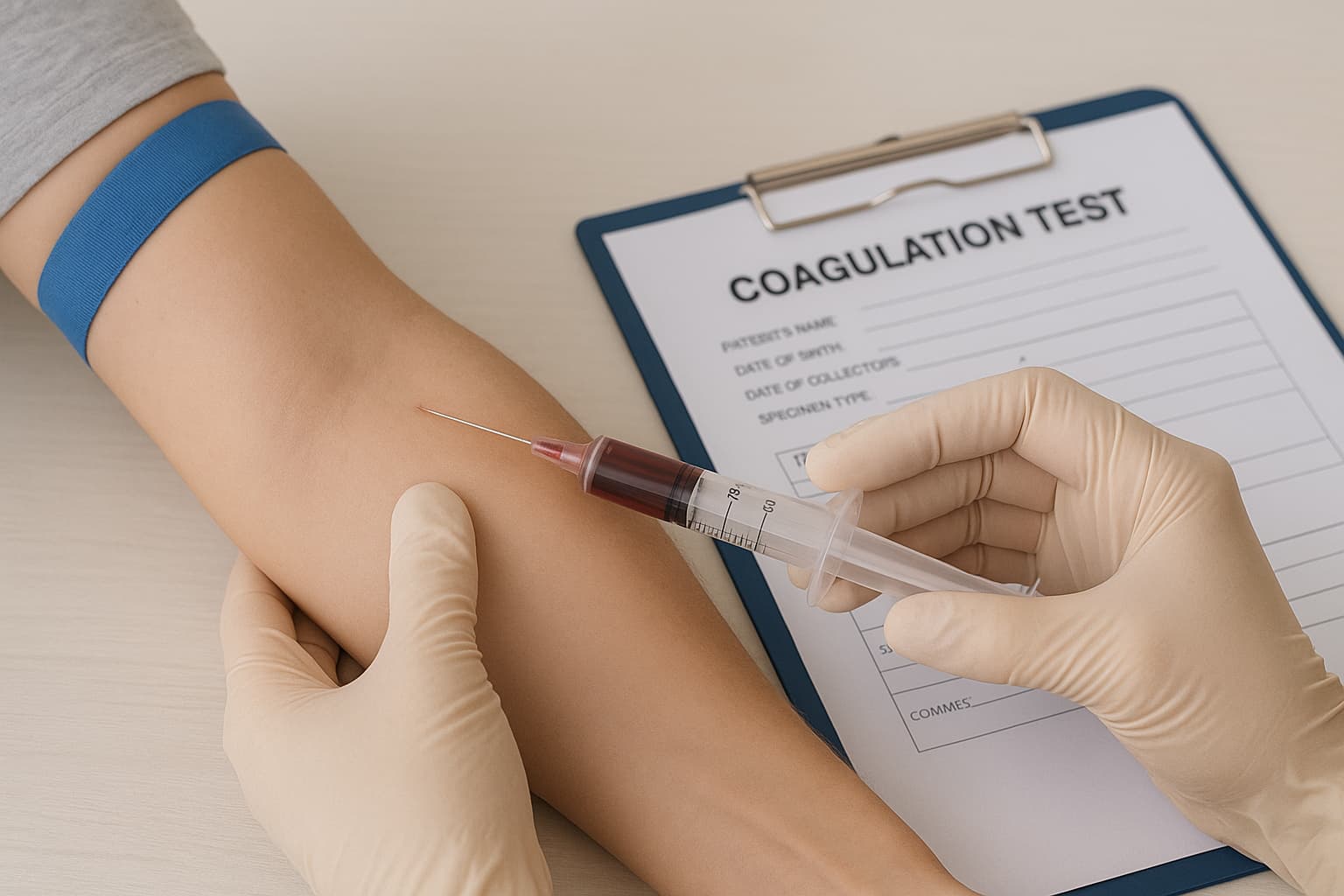
Blood remains fluid within the vascular system yet must solidify promptly when a vessel is injured. When either direction fails, patients can face severe bleeding to life-threatening embolic events. That said, a coagulogram test is a structured way to evaluate these pathways and pinpoint where the imbalance lies.
A modern coagulation workup extends beyond a simple blood clot test. It now functions as an integrated set of coagulation studies designed to evaluate clotting factor activity, platelet behaviour, fibrin formation, inhibitor presence, anticoagulant influence, and liver-derived protein synthesis.
For pathologists, the coagulogram is central to the diagnosis of bleeding disorders, perioperative risk assessment, and monitoring of antithrombotic therapy.
Why the Coagulogram Test Matters
Abnormal clot formation can occur without injury, producing thrombi that migrate to the lungs, brain, or coronary arteries. Conversely, inadequate clot formation produces prolonged bleeding even after minor trauma. A structured coagulation profile test brings clarity to both scenarios by quantifying reaction times, factor levels, and platelet adequacy.
Major clinical triggers for ordering a coagulation screening test include,
- Unexplained bleeding from gums, nose, or surgical sites.
- Excessive bruising or petechial eruptions.
- Suspected DIC, inherited factor deficiencies, or acquired liver dysfunction.
- Monitoring of blood-thinner therapies, e.g., warfarin and heparin therapy.
- Pre-operative screening prior to invasive procedures.
- Evaluation of thrombophilia in patients with unexplained DVT or pulmonary embolism.
Beyond diagnosis, these tests indicate whether clotting factors are reduced, absent, or overexpressed. They also give an indirect look at hepatic synthetic function.
Clinical Conditions Flagged
A complete blood coagulation test identifies patterns suggestive of multiple systemic disorders.
Bleeding Tendencies
- Vitamin K deficiency.
- Liver disease.
- Fibrinogen abnormalities.
- Immune-mediated factor inhibition.
- Placental abruption.
- Inherited single-factor deficiencies, e.g., haemophilia.
Excessive Clotting
- Autoimmune disorders, including lupus.
- Malignancy.
- Prolonged immobility.
- Infections such as sepsis or COVID-19.
- Deficiency of vitamins B6, B12, and folate.
- Myeloproliferative states.
Clinical symptoms can range from heavy menstrual bleeding to hemarthrosis or unexplained swelling of limbs due to DVT. Shortness of breath, chest pain, and sudden collapse raise suspicion for pulmonary embolism.
The Coagulation Profile Test List
A standard coagulogram includes multiple assays. Many laboratories expand the panel to include platelet parameters and thrombin-fibrinogen interactions.
Coagulation Profile Components
| Test | Measures / Purpose | Abnormal Findings Suggest |
|---|---|---|
| CBC | Platelets, RBCs, WBCs | Low platelets, anaemia influencing clotting |
| PT / INR | Extrinsic pathway, vitamin K–dependent factors | Warfarin effect, hepatic dysfunction, vitamin K deficiency |
| aPTT | Intrinsic pathway activity | Factor deficiency, intrinsic pathway inhibitors |
| Fibrinogen (Factor I) | Fibrinogen concentration | Bleeding, DIC, placental abruption |
| Factor V Assay | Factor V levels | Liver disease, fibrinolysis, DIC |
| Platelet Count | Circulating platelet volume | Leukaemia-related reduction or elevation in myeloproliferative states |
| Thrombin Time | Conversion of fibrinogen to fibrin | Cancer, liver disease, dysfibrinogenemia |
| Bleeding Time | Small-vessel clotting mechanics | Platelet dysfunction or vessel wall abnormalities |
Test Procedure and Sampling
A coagulation time measurement depends heavily on specimen integrity. Errors in blood collection methods or anticoagulant ratios lead to misleading outputs.
Venipuncture and Collection
- Standard venipuncture from an antecubital or dorsal hand vein.
- Blood collected into 3.2% sodium citrate tubes.
- The correct fill line is essential to retain a 1:9 anticoagulant-to-blood ratio.
- Mild inversion to avoid clotting without activating platelets.
No fasting is required. However, clinicians should review medication regimens, particularly antiplatelet agents and anticoagulants. Pausing therapy may also be necessary.
Turnaround Time
Most laboratories report results by the next day, though urgent DIC panels may be expedited. Automated assays performed on a coagulation analyser enhance reproducibility and shorten the turnaround time.
Understanding the Results
Values are reported as seconds to clot formation, or as protein concentrations. Interpretation depends on:
- Lab-specific coagulation profile normal value ranges
- Pattern of PT/INR and aPTT extension or contraction
- Clinical presentation
- Medication profile
- Factor assays
- Platelet function tests
Low Levels or Absence of Clotting Factors
Reduced factor levels signal a bleeding tendency. These reductions may involve a single factor (frequently inherited) or multiple factors (commonly acquired due to liver disease, DIC, or vitamin K deficiency).
Increased Clotting Factor Levels
Higher-than-expected factor levels suggest a pro-thrombotic state, particularly in inflammatory or neoplastic conditions. Such individuals may need adjustments to hormone therapy, lifestyle modifications, or preventive anticoagulation.
Half-Life of Coagulation Factors
Knowledge of factor half-life supports the timing of repeat assays and therapeutic monitoring. For instance, Factor VII has a short half-life, making PT/INR sensitive to acute vitamin K changes, whereas fibrinogen and Factor II decline more slowly.
Additional Points for Pathology Practice
Normal Coagulation Profile Does Not Exclude Disease: Isolated platelet dysfunction, mild factor deficiencies, or early DIC may appear normal on initial screening. Clinical correlation remains essential.
Repeated Sampling: Gradual shifts in coagulation time or fibrinogen concentration reveal early progression of systemic disease, especially hepatic failure or sepsis.
Expanding the Workup: In ambiguous cases, specialised tests such as mixing studies, lupus anticoagulant panels, anti-Xa assays, or specific inhibitor quantification refine the diagnosis.
Conclusion
A coagulogram test is an organised method to evaluate clot formation, factor adequacy, platelet integrity, and potential thrombotic risk. When interpreted alongside symptoms and clinical history, it directs diagnostic decisions, guides therapy choices, and supports safe surgical planning.
Whether assessing unexplained bleeding, evaluating thrombophilia, or monitoring anticoagulant therapy, a well-performed coagulation profile test continues to anchor laboratory decision-making in haematology and pathology.
Also check - Fibrinogen Test: Why It’s Done and What Results Mean
Get Started at ₹1!
Try Flabs for a full month for just ₹1.
Follow us on




