
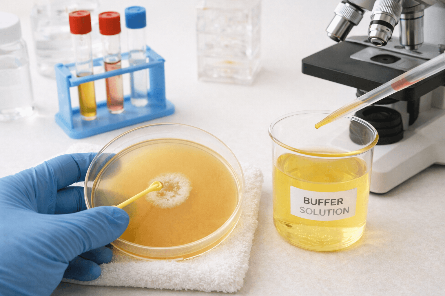
Laboratories rely on straightforward biochemical assays to reveal metabolic traits that distinguish one microorganism from another. The hydrolysis test family gives pathologists and microbiologists a window into exoenzyme activity, substrate degradation patterns, and species-level variability.
As antimicrobial resistance grows and faster reporting becomes essential, renewed attention on classical assays feels warranted. The hydrolysis test remains relevant, practical, and adaptable across routine diagnostic labs, teaching labs, and automated systems.
What the Hydrolysis Test Evaluates
A hydrolysis test, and every variant of it, examines the ability of microorganisms to secrete enzymes that degrade complex substrates into smaller, functional units.
Because large macromolecules cannot pass through cell membranes, organisms depend on extracellular enzymes such as amylase, gelatinase, caseinase, urease, and DNase. When present, these enzymes cleave polymers, forming visible changes in culture media.
Common formats include:
- Starch hydrolysis test
- Casein hydrolysis test
- Gelatin hydrolysis test
- Urea hydrolysis test
- Hippurate hydrolysis test
- DNA hydrolysis DNase test, including the DNA hydrolysis test for Staphylococcus aureus
Each assay has its own interpretation rules, applications, and quality indicators; the starch hydrolysis test remains one of the best-known models for teaching the principle.
Principle of Starch Hydrolysis Test
Starch contains amylose and amylopectin, forming a high-molecular-weight polysaccharide. Because starch cannot diffuse across bacterial membranes, only organisms that secrete extracellular amylase can break it down.
The principle of the starch hydrolysis test hinges on detecting starch degradation using iodine, which stains intact starch blue-black. Any hydrolysed area remains clear, forming a translucent halo surrounding amylase-producing colonies.
Such behaviour differentiates species in genera like Bacillus, Clostridium, Corynebacterium, Streptococcus, Enterococcus, Fusobacterium, and Pseudomonas.
Media, Reagents, and Equipment
Media Used
- Starch agar
- Mueller–Hinton agar with starch
- Heart Infusion Agar with 2% starch
Preparation involves dissolving dehydrated media in water, melting the mixture, sterilising at 121°C for 15 minutes, and then pouring plates at roughly 45°C.
Reagent: Gram’s iodine, prepared from iodine, potassium iodide, and optional sodium bicarbonate, highlights the distribution of residual starch.
Equipment: Petri dishes, autoclave, incubator, inoculating loop, Bunsen burner, dropper, weighing scale.
Controls
Positive: Bacillus cereus, Streptococcus bovis
Negative: Escherichia coli, Staphylococcus aureus
Starch Hydrolysis Test Procedure
- Inoculate fresh colonies onto starch agar using a straight-line streak.
- Incubate at 35 ± 2°C for 48 hours.
- Add iodine gently across the plate surface.
- Observe colour distribution.
Interpretation
Positive: A clear halo appears around colonies while the remainder of the plate turns blue-black.
Negative: No halo forms; the entire medium stains uniformly.
Species like B. subtilis, B. cereus, B. megaterium, Clostridium perfringens, and S. bovis hydrolyse starch.
Non-hydrolysers include Corynebacterium diphtheriae, C. difficile, C. botulinum, viridans streptococci (except S. bovis), E. coli, S. aureus, and Pseudomonas aeruginosa.
Precautions
- Apply iodine only after completing 48 hours of incubation.
- Keep streaks at least 25 millimetres apart.
- Avoid high-glucose media that may shift microbial metabolism.
- Read plates promptly since iodine colour fades with time.
Quality control: B. cereus and S. bovis consistently produce halos, while E. coli and S. aureus remain negative.
Other Hydrolysis Tests in Microbiology
| Hydrolysis Test | Enzyme Detected | Primary Use |
|---|---|---|
| Starch hydrolysis test | Amylase | Differentiation within Bacillus, Streptococcus, and Clostridium |
| Casein hydrolysis test | Caseinase | Evaluation of proteolytic activity; the casein hydrolysis test principle aligns with detecting clear zones in skim-milk agar |
| Gelatin hydrolysis test | Gelatinase | Assessment of protein degradation requires gelatin hydrolysis test reagent to visualise liquefaction |
| Urea hydrolysis test | Urease | Identification of urease-producing Enterobacteriaceae |
| Hippurate hydrolysis test | Hippuricase | Distinguishing Campylobacter jejuni and Streptococcus agalactiae |
| DNA hydrolysis DNase test | DNase | Identifying S. aureus and other DNase-positive organisms |
Each assay follows the broader logic of substrate breakdown, producing visible change. The casein hydrolysis test procedure and gelatin hydrolysis test workflows mirror the starch assay concept, although indicators and media differ.
Applications in Microbiology
Hydrolysis assays deliver meaningful insights into metabolic diversity.
1. Species Differentiation
The starch hydrolysis test separates S. bovis from bile-esculin-positive viridans streptococci. It also supports the separation of Chryseobacterium indologenes (positive) from Elizabethkingia meningoseptica (negative).
Casein and gelatin assays add clarity for proteolytic organisms, especially in environmental and wound isolates.
2. Rapid Recognition of Enzyme-Producing Pathogens
The DNA hydrolysis test for Staphylococcus aureus remains a trusted adjunct in staphylococcal workflows. DNase activity correlates strongly with pathogenicity markers.
3. Integrating Biochemical Tests into Digital Systems
Modern LIS platforms incorporate structured test panels, automated reporting, and media-linked interpretation templates. Hydrolysis tests transition smoothly into such systems, enhancing turnaround time without changing laboratory methodology.
4. Teaching Exoenzyme Biology
Students gain a direct view of enzyme secretion and substrate degradation. Since amylase production varies among species, observing differences in halo size enriches microbial profiling.
Amylase Biology in Context
Amylase is synthesised intracellularly and secreted externally once the amylase gene is expressed. It cleaves starch into glucose and maltose, which microorganisms transport for energy. Strong producers may hydrolyse starch beyond the immediate growth zone, forming wide halos. Such variability supports species differentiation.
Limitations
- Not meant as a standalone confirmation test; biochemical and molecular panels add higher specificity.
- Plates treated with iodine cannot be subcultured.
- Requires extended incubation.
Integrating with Flabs Pathology Software
Adopting biochemical assays becomes significantly easier when paired with a digital workflow that reduces manual tracking. Flabs Pathology Software, an AI-powered LIS platform, brings together patient registration, automated analysis pipelines, and QR-coded report delivery.
Microbiology teams recording hydrolysis test outcomes gain structured templates, standardised result fields, and smoother interpretation layers.
A few capabilities align neatly with hydrolysis workflows.
- Streamlined operations: Enter observations for starch or casein assays directly into customizable panels, generate reports instantly, and share results through WhatsApp integration.
- Enhanced patient experience: Patients access hydrolysis-related reports from a secure phone-number login, avoiding repeated inquiry calls.
- User-friendly design: Technicians migrate from paper-based worksheets to an intuitive interface designed for rapid entry of enzyme assay outcomes.
- Scalable architecture: Expand to multiple sections or collection points without reorganising software infrastructure.Secure data handling: Encrypted storage, disaster recovery, and controlled user access safeguard microbiology records.
Labs adopting hydrolysis tests gain smoother throughput once Flabs centralises processes.
You can explore these features through a 5-day free trial by submitting your details. The platform adapts to small labs and larger diagnostic centres with equal ease.
Conclusion
Hydrolysis tests are important in microbial enzyme profiling. The starch hydrolysis test procedure anchors the concept through its clear visual endpoint and stable interpretation rules.
When paired with digital tools such as Flabs LIS, a lot becomes easier for pathologists and healthcare professionals. Integrating biochemical assays into a modern LIS environment supports accuracy and operational clarity from sample to report.
Also check - CMV Avidity Test: What It Measures and Why It Matters
Get Started at ₹1!
Try Flabs for a full month for just ₹1.
Follow us on




