
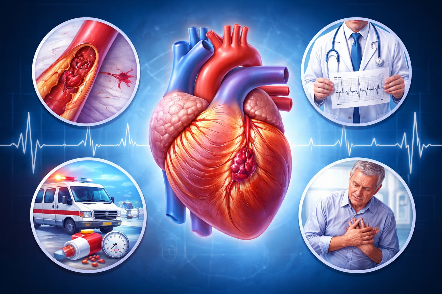
You’re staring at a set of cardiac enzymes and an ECG strip. The diagnosis is myocardial infarction. But what type is it? For pathologists and clinicians, that distinction is not just academic. It is related to proper diagnosis.
It changes how you interpret the slide, how you advise your colleagues, and how you understand what happened to the patient. Knowing the classification is mandatory for many more reasons. So, let’s get straight to the types of myocardial infarction.
What is Myocardial Infarction
The myocardial infarction definition is straightforward: it’s the death of heart muscle cells (myocytes) due to prolonged ischemia.
That ischemia usually stems from a mismatch between oxygen supply and demand. But how that mismatch happens, that’s where the classification gains attention.
The Five Main Types of Myocardial Infarction
The universal classification system, backed by cardiology and pathology societies, splits types of myocardial infarction into five distinct categories.
| Type of Myocardial Infarction | Mechanism | Clinical Situation | Diagnostic Clues |
|---|---|---|---|
| Type 1 MI | Atherosclerotic plaque rupture leads to thrombus formation and coronary artery blockage. | Primary coronary artery event caused by unstable plaque. | Includes STEMI (complete occlusion with ST elevation) and NSTEMI (partial occlusion without ST elevation). Elevated troponin and ischemic ECG changes. |
| Type 2 MI | Imbalance between myocardial oxygen supply and demand without acute coronary thrombosis. | Severe anemia, sepsis, tachyarrhythmia, hypotension, or respiratory failure. | Troponin elevation with clinical evidence of ischemia but no plaque rupture. ECG changes may vary. |
| Type 3 MI | Sudden cardiac death due to suspected myocardial ischemia before biomarkers can be measured. | Cardiac arrest occurring before hospital evaluation or laboratory testing. | Ischemic symptoms or ECG changes observed before death. Biomarker confirmation unavailable. |
| Type 4 MI | Infarction related to percutaneous coronary intervention (PCI) procedures. | Complications during or after PCI, including vessel injury or stent problems. | Type 4a: procedure-related myocardial injury. Type 4b: infarction caused by stent thrombosis. |
| Type 5 MI | Myocardial injury associated with coronary artery bypass graft (CABG) surgery. | Occurs during or shortly after bypass surgery. | Marked elevation of cardiac biomarkers with supporting ECG or imaging evidence. |
Type 1: (STEMI and NSTEMI)
Type 1 is the most common type of myocardial infarction you’ll encounter.
It’s the one that fits the classic image: an unstable atherosclerotic plaque ruptures, triggering platelet aggregation and a cascade that ends in a thrombus. The coronary artery is suddenly occluded.
When you’re looking at tissue, you’re searching for evidence of that acute event. The causes of myocardial infarction in this case are purely atherothrombotic. The patient’s myocardial infarction symptoms were likely sudden and severe.
For example, crushing chest pain, diaphoresis, and shortness of breath.
The myocardial infarction ECG will often show ST-segment elevations (STEMI) or new left bundle branch blocks, though it can present as a non-ST elevation MI (NSTEMI) as well.
Why does this matter to you? Because Type 1 tells a story of progressive vascular disease. It’s the wake-up call for the patient and the definitive endpoint for the plaque.
Type 2: The Imbalance
In Type 2, there is no plaque rupture. Instead, you have a fixed stenosis or normal arteries. But the heart is starved due to external factors. Think of it as a supply-and-demand equation gone wrong.
A patient with sepsis, profound hypotension, or rapid atrial fibrillation who also has elevated troponin. That’s Type 2. The oxygen supply drops, or the demand spikes (or both), and the heart muscle suffers.
The types of myocardial infarction type 1 scenarios are mechanical; Type 2 is physiological. As a pathologist, you might see diffuse subendocardial injury rather than the focal, full-thickness necrosis typical of Type 1.
The ECG might show diffuse ST depressions rather than localised elevations.
The distinction here is vital because treating the underlying cause (e.g., stabilising blood pressure) is the primary move, not just opening an artery.
Type 3: The Silent Witness
Type 3 is the pathologist's domain. This diagnosis is made post-mortem. A patient dies suddenly with symptoms suggestive of myocardial ischemia, and maybe they had new ST elevations or a presumed new LBBB before they were arrested. But they died before blood could be drawn for troponins.
You are the one who confirms it. The autopsy reveals a recent myocardial infarction or a thrombosed artery. This classification acknowledges that MI can be fatal before we get the lab confirmation we usually rely on.
Type 4: Associated with PCI
Coronary intervention saves lives, but it's not without myocardial consequences. In other words, Type 4 is associated with percutaneous coronary intervention (PCI). It splits into two subcategories.
Type 4a: MI occurring within 48 hours of percutaneous coronary intervention. With normal baseline troponin, you'll see elevation to more than 5x the 99th percentile URL. With elevated but stable or falling baseline levels, you need a rise of at least 20 percent.
Type 4b: Stent thrombosis-related MI. Documented thrombosis by angiography or autopsy, accompanied by ischemic symptoms or ECG changes.
Type 5: Associated with CABG
Coronary artery bypass grafting (CABG) places unique demands on the myocardium.
Type 5 MI occurs within 48 hours of CABG, with troponin elevation exceeding ten times the 99th percentile URL, plus new pathological Q waves, new graft occlusion, or imaging evidence of new viable myocardium loss.
The Anatomical Picture: Beyond the Numbers
While the universal classification is based on mechanism, the location matters too. You might hear a clinician mention anteroseptal myocardial infarction. What are they telling you?
They’re localising the damage. Anteroseptal refers to the front wall of the heart and the septum dividing the ventricles. This usually points to a blockage in the left anterior descending (LAD) artery. The ECG changes tell the clinician where to look. For you, in the lab, it tells you which part of the heart to examine most closely for reperfusion injury or scarring.
ECG and the Types: Connecting the Dots
You can’t talk about types of myocardial infarction and ECG changes without linking them to the classification. The ECG is your bedside map.
- Type 1 STEMI: You’ll see that tombstone ST elevation. It screams, "Open me now!"
- Type 1 NSTEMI: You might see ST depression or T-wave inversions. The artery is not fully occluded, but it's critically unstable.
- Type 2: The ECG is often variable. It may show diffuse changes or simply tachycardia without localizing signs. The story is in the chart, not just the tracing.
Why Do These Distinctions Matter for Treatment?
The classification directly dictates myocardial infarction treatment. You can't treat all heart attacks the same way.
- For a Type 1 MI, the goal is rapid reperfusion. It involves either with angioplasty/stenting or clot-busting drugs.
- For a Type 2 MI, the focus shifts to correcting the imbalance. Treat the sepsis. Slow the heart rate. Raise the blood pressure. Stenting a clean artery won't help someone who is in shock.
- For a Type 4 or 5 MI, the treatment is often supportive and focused on the procedure itself.
The Root Causes of MI
The causes of myocardial infarction vary by type, but they all trace back to the health of the arteries and the blood.
Lifestyle and Dietary Factors
Poor Diet: High intake of saturated fats, trans fats, cholesterol, processed foods, sugar, and red meat promotes plaque buildup in arteries.
Obesity and Physical Inactivity: Excess body weight and lack of exercise increase blood pressure, cholesterol levels, and cardiovascular risk.
Excessive Alcohol: Heavy drinking can raise blood pressure and cholesterol, increasing the risk of coronary artery disease.
Clinical Risk Factors
High Blood Pressure (Hypertension): Damages arterial walls and promotes plaque formation.
High Cholesterol: Elevated LDL (“bad”) cholesterol contributes to plaque buildup, while HDL (“good”) cholesterol helps remove it.
Diabetes Mellitus: High blood glucose damages blood vessels and increases cardiovascular risk.
Smoking: Causes vascular damage, increases blood pressure, and promotes clot formation.
Genetic and Autoimmune Factors
Genetic Predisposition: Family history of heart disease and inherited conditions such as familial hypercholesterolemia increase the likelihood of atherosclerosis and MI.
Autoimmune Diseases: Conditions like lupus and rheumatoid arthritis cause chronic inflammation that may accelerate plaque formation.
Infectious and Environmental Factors
Infections: Certain viral or bacterial infections may cause inflammation in coronary arteries, increasing plaque instability. Myocarditis can also contribute to cardiac complications.
Air Pollution: Long-term exposure to fine particulate matter can cause vascular inflammation and increase cardiovascular risk.
Demographic and Psychological Factors
Age: Risk increases with age, especially after 45 years in men and 55 years in women.
Gender: Men develop MI earlier than women, while women’s risk rises after menopause due to reduced estrogen protection.
Stress: Chronic stress, anxiety, and depression may increase blood pressure and inflammatory responses, contributing to heart disease.
For Type 1, it's the culmination of years of plaque buildup, driven by hypertension, hyperlipidemia, smoking, and diabetes.
For Type 2, the causes are broader: anaemia, respiratory failure, hypotension, and extreme hypertension. You’re looking at systemic illness damaging the heart secondarily.
Complications: What Comes Next?
No matter the type, complications of myocardial infarction follow a predictable, grim timeline. Recognizing them is part of your job.
Arrhythmias: Damaged conduction pathways lead to chaos.
Heart Failure: When the pump is broken, fluid backs up.
Myocardial Rupture: A rare but catastrophic event days after a transmural infarct, where the necrotic wall gives way.
Pericarditis: Inflammation of the sac surrounding the heart (Dressler's syndrome) can occur weeks later.
Ventricular Aneurysm: Scar tissue bulges with each contraction.
For the pathologist, these complications are the final chapter in the story of the acute myocardial infarction.
Wrapping Up
So, next time you’re reviewing a case, walk through the types of myocardial infarction in your mind.
Was it a spontaneous plaque rupture (Type 1)? Or was it a demand issue (Type 2)?
Did it happen during a procedure (Type 4 or 5)?
Your ability to distinguish these is about piecing together the pathophysiology that led that patient to your lab or your table.
The classification exists to enable precision for pathologists. It turns a "heart attack" into a specific diagnosis with specific implications. And that, ultimately, is what the medical field needs.
Also check:
Get Started at ₹1!
Try Flabs for a full month for just ₹1.
Follow us on




