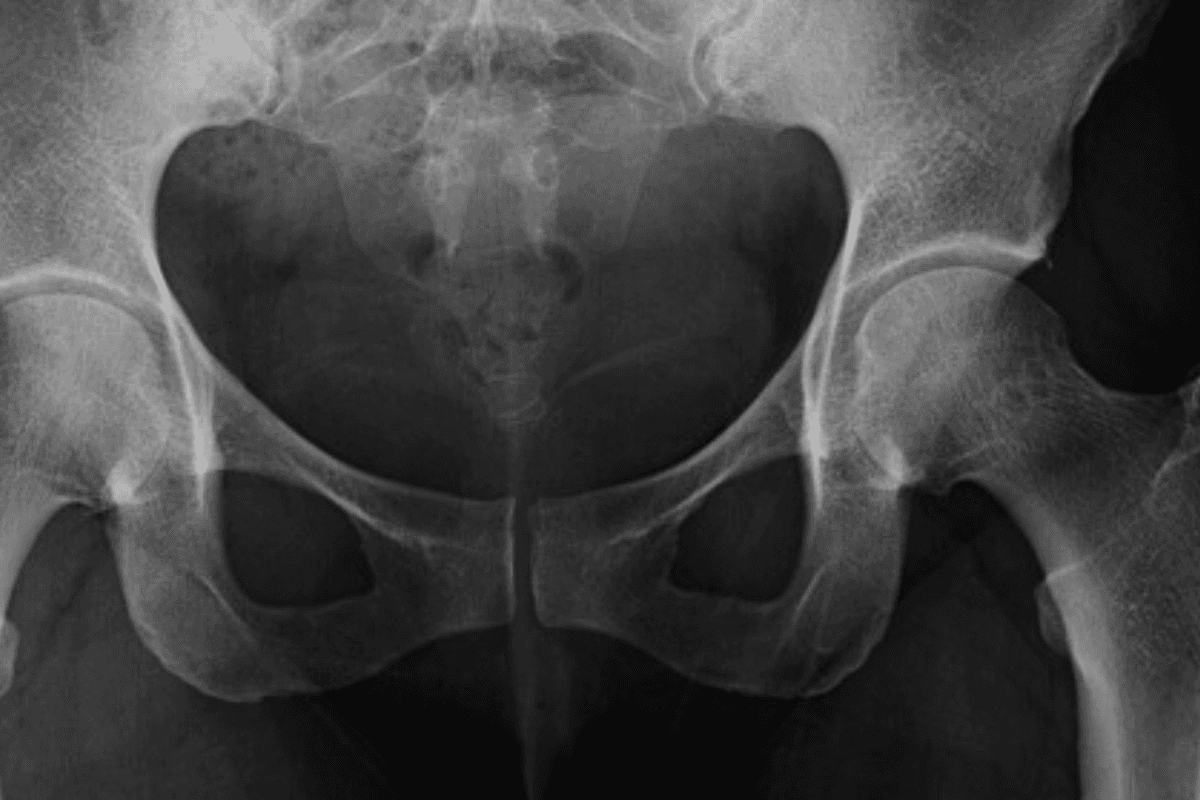
Coccyx X-Ray is performed to assess the condition of the human tailbone, when a patient reports sustained pain of the tailbone, due to accidents, injuries or bone-related conditions. Based on the outcomes, the cause and level of damage to the tailbone may be detected. Some conditions like Sacroiliac joint dysfunction also mimic symptoms related to Coccyx pain, when your doctor may request to undergo the procedure.
To assess Coccyx pain completely, X-Ray is taken in different views (Anteroposterior and Lateral). A dynamic X-Ray when a person is sitting or standing is required to assess the actual pain and stress on the tailbone. So, when do you need a Coccyx X-Ray? How is it performed? What do results mean? Let’s find answers to these questions in this discussion.
What is Coccyx
Coccyx is the tailbone, situated at the bottom of your spine. It is a small, curved bone that most people may not know exists, unless they fall on and injure it. But it supports you throughout when you’re sitting or standing, by connecting many muscles and ligaments. It is like one leg of a tripod that distributes your body weight evenly and keeps you stable when you’re sitting. It is often referred to as a single bone, when it is a combination of several vertebrae fused together.
Indications for a Coccyx X-Ray
- A Coccyx X Ray may be requested in the following cases:
- Trauma or Injury: Any injury or trauma of Coccyx due to direct fall onto the tailbone, sports related injuries, vehicular accidents, pregnancy, overweight, etc.
- Coccydynia: Persistent tailbone pain lasting more than a few weeks, or a pain of Coccyx that doesn’t respond to conservative treatments might necessitate a Coccyx X-ray.
- Suspected fracture or dislocation of the coccyx, or Persistent pain while sitting, especially on hard surfaces.
- Abnormal growth or mass in the coccygeal region.
- Pain during bowel movements or sexual intercourse.
- Suspected osteomyelitis (bone infection) of the coccyx.
- Ruling out other conditions that may mimic coccyx pain (e.g., sacroiliac joint dysfunction).
- Some types of cancers are Chordoma, Prostate cancer, or Colorectal (colon) cancer can also damage Coccyx, requiring an X-Ray to diagnose.
It's important to note that while X-rays can provide valuable information about the bony structures of the coccyx, they may not always reveal soft tissue problems. In some cases, additional imaging studies like MRI or CT scans may be necessary for a comprehensive evaluation.
The Procedure: What to Expect
Preparation
Before the Coccyx X-Ray, there are no general preparatory steps needed. However, sometimes, the urinary bladder should be empty in AP Coccyx X-Ray for best results. Your doctor might also advise about cleaning enema and gas in the colon. Inform any recent illnesses, medical conditions, or ongoing medications. Mention if you’re pregnant or expecting to be.
Remove all metal objects, jewellery, and electronic devices from your body. You might be asked to change into a hospital gown to avoid interference from clothing with metal fasteners. A thread in clothes may be an obstruction. Shielding may be possible for males, not females.
During X-Ray
Coccyx X-Ray Positioning
Lateral View: Lying on Your Side
It is also known as sacrum and coccyx lateral view, which demonstrates the most distal region of the spine. It is commonly used in conjunction with AP view, but can also be used as a solo projection. Lateral Coccyx X-Ray is the best to investigate both acute and chronic sacral and coccyx pain.
AP (anteroposterior) View: Lying on Your Back
AP view helps to visualise the pathology of Coccyx, especially fractures. Emptying bladder prevents superimposition of structures over the tailbone region. As mentioned, Coccyx x-ray AP view is sometimes combined with Lateral view for better insights.
Sitting Position: To Assess the Coccyx under Stress
For a dynamic X-ray, the radiologist takes two images of your coccyx: one while standing and another while sitting. Comparing these images helps determine if your coccyx moves excessively when you sit, which could indicate instability. It allows for a precise diagnosis in about 70% of cases.
The doctor might also examine your lower back to rule out referred pain not caused by the coccyx. This examination involves bending over, lying down, lifting each leg, and checking your reflexes.
Proper patient and part conditioning is essential in Coccyx X-ray positioning. During the procedure, the patient's face should be upward in AP and lateral Coccyx X-Ray. You will be given a pillow for head support and legs are stretched. Pelvis should not be in a rotated position. Also, the central ray, collimation, and respiration should be properly aligned and managed.
Post-Procedure
There are no precautions to take after Coccyx X-Ray. You can resume your normal activities. As such, there is no recovery time. Radio-exposure is not significant to have side effects, although you must discuss any concerns with your doctor.
Your consultant radiologist will view images and forward it to the referring physician. Results from the X-Ray are discussed and further tests may be suggested if there are any abnormalities. Store the X-ray images safely if they are given to you, as they may be needed
A Normal Coccyx X-Ray
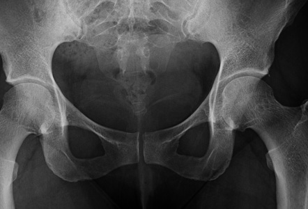
Coccyx X-Ray AP View
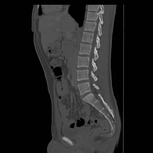
Coccyx X-Ray Lateral View
Interpreting Coccyx X-Ray Results
Results of Coccyx X-Ray are analysed and interpreted by expert radiologists or orthopaedic specialists. They look for various factors like alignment, bone density, fractures, joint spaces, presence of foreign bodies, soft tissue shadows, and surrounding structures. Based on the conditions of various structures, they produce a diagnosis.
- A healthy normal Coccyx X-Ray shows a natural, slightly curved line of Coccyx. If there are any deviations or angulations, it calls for attention indicating a dislocation or fracture.
- Any breaks or discontinuities in the bone structure, whether as hairline or complete, indicate fractures.
- Abnormal bone density in the Coccyx X-ray might be indicative of osteoporosis or bone tumours.
- The spaces between coccygeal segments should be uniform. Irregularities might indicate arthritis or other joint issues.
- Coccyx X-ray can also suggest any soft-tissue issues in a subtle way. For further validation, a CT or MRI will be suggested in case of suspicion.
- The relationship between Coccyx to the scrotum or pelvic bones is also evaluated from Coccyx X-Ray imaging.
Of course, no interpretation is complete without considering the clinical history and symptoms of patients. Based on these additional factors, a complete diagnosis is produced. If necessary, further tests may be suggested to detect the exact problem.
Diagnosing Coccydynia
Coccydynia is a medical term for tailbone pain, derived from (“coccyx” = tailbone, “dynia” = pain). It is a tailbone pain that can be discomforting, caused due to injuries or related health conditions. When diagnosing Coccydynia, Coccyx X-Ray uses a dynamic method of X-Ray, which images when the patient is sitting and standing. That is, it considers medical imaging under actual stress conditions and how your Coccyx is responding to them.
X-Rays from two images are compared to detect pelvic rotation angle and change in position of the Coccyx from sitting to standing. If these are outside of range (between 5-25 degrees) or too much or too little Coccygeal movement describes the cause of pain.
Conclusion
Coccyx X-Ray is an important diagnostic step in detecting many problems along with simple tailbone pain. A tailbone pain is usually not problematic, often resolving itself with time, without external intervention. But it can also indicate osteoporosis, and other bone and joint related problems. If you have sustained pain, you might wanna consult your physician for it.
Get Started at ₹1!
Try Flabs for a full month for just ₹1.
Follow us on