An IHC stainer automates key steps in immunohistochemistry, helping histopathology laboratories standardize staining while reducing repetitive manual work. For labs processing increasing numbers of tissue slides, choosing the right automated IHC stainer is not simply about buying the machine with the highest slide capacity.
The laboratory must also consider antibody compatibility, reagent systems, staining protocols, quality controls, throughput, turnaround time, service support and how the instrument fits into the wider histopathology workflow.
Current platforms illustrate how differently IHC systems are designed. Biocare's ONCORE Pro processes up to 36 slides and supports IHC, ISH, FISH and CISH, while Roche's BenchMark ULTRA provides 30 independently controlled slide positions. Agilent's Dako Omnis offers a 60-slide capacity and supports IHC and ISH workflows with laboratory connectivity.
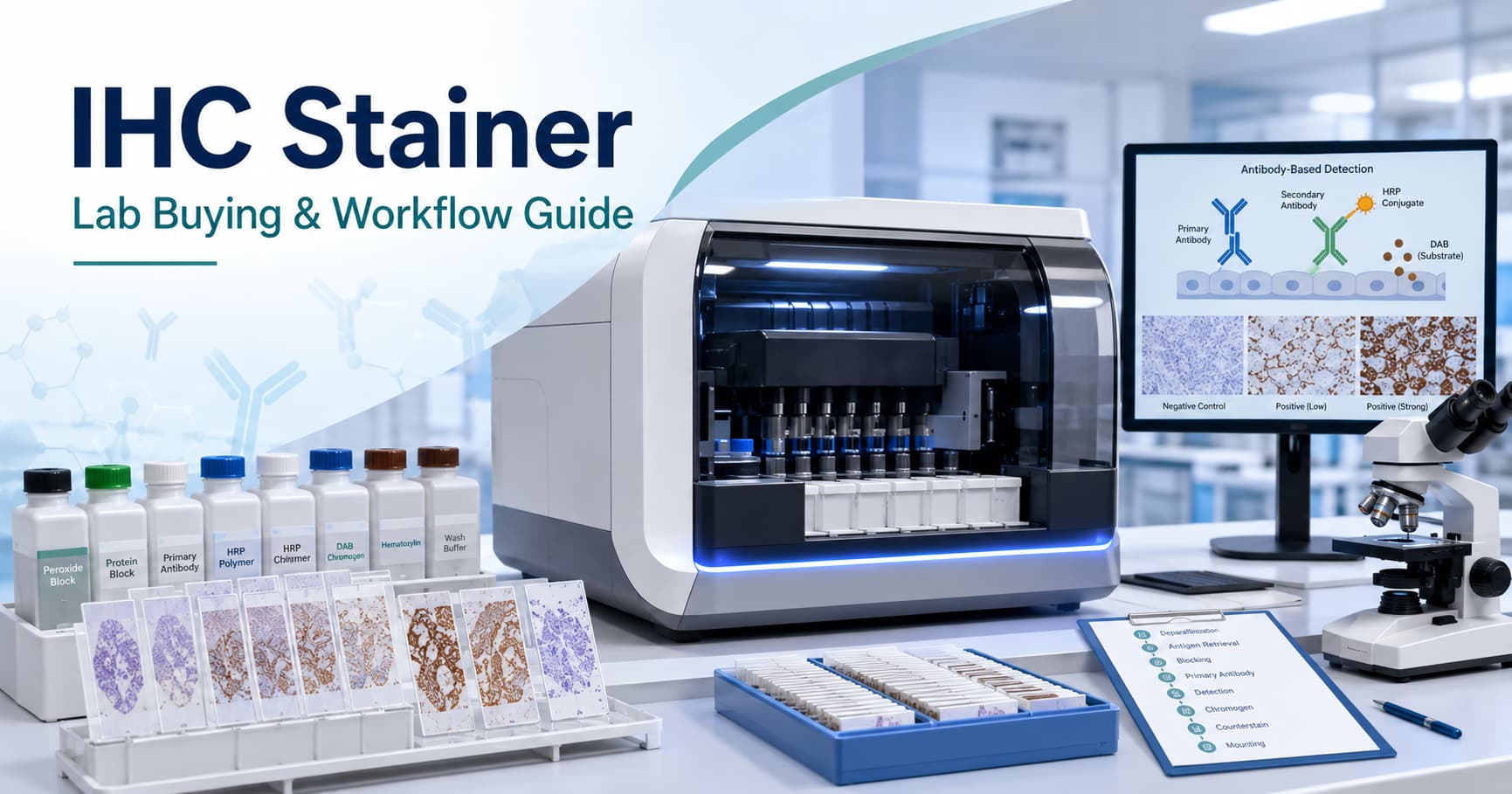
Image suggestion: Automated IHC stainer with slide positions, reagent containers and histopathology tissue slides.
What Is an IHC Stainer?
An IHC instrument or IHC stainer is a laboratory system used to perform some or most of the immunohistochemical staining workflow on prepared tissue slides.
Immunohistochemistry uses antibodies to identify specific antigens or proteins within tissue while preserving their location within the tissue architecture. Antibody binding is then visualized through a chromogenic or fluorescent detection method.
Automated systems can standardize steps such as reagent application, incubation, washing, antigen retrieval, detection and counterstaining. The exact level of automation varies by platform.
For laboratories reviewing their broader histopathology setup, FLABS also provides a guide to the pathology lab equipment list.
IHC Staining Principle
The IHC staining principle is based on specific binding between an antibody and its target antigen in a tissue section.
After the primary antibody binds to the target, a detection system creates a visible signal. In chromogenic IHC, enzymes such as horseradish peroxidase can react with a chromogen to create a coloured deposit at the antigen site. Fluorescent methods instead use fluorophores that can be viewed with fluorescence microscopy.
In routine diagnostic histopathology, the final staining pattern is assessed by a qualified pathologist together with tissue morphology, controls and other clinical findings.
IHC Staining Procedure
The exact IHC staining procedure depends on the antibody, tissue, detection system and validated laboratory protocol, but a typical workflow includes:
- Fixation and tissue processing to preserve tissue morphology.
- Sectioning and slide preparation from paraffin-embedded tissue.
- Deparaffinization and rehydration.
- Antigen retrieval where required to expose antibody-binding sites.
- Blocking to reduce unwanted background staining.
- Primary antibody application.
- Detection and visualization, commonly through enzyme/chromogen-based systems.
- Counterstaining, often with haematoxylin.
- Review of controls and stained tissue before interpretation.
Automated systems may perform many of these stages within one instrument, but labs should confirm exactly what is automated on the platform they are evaluating. Roche's BenchMark ULTRA, for example, automates baking, deparaffinization, conditioning and staining.
Image suggestion: Simple IHC workflow infographic: Tissue → Antigen Retrieval → Primary Antibody → Detection → Chromogen → Pathologist Review.
What Is IHC Staining Used For?
IHC is widely used in diagnostic pathology to help:
- Characterize and classify tumours
- Determine tissue or cell lineage
- Distinguish morphologically similar lesions
- Assess selected prognostic or predictive biomarkers
- Support evaluation of hormone-receptor or therapeutic-marker status
- Identify selected infectious agents or proteins in tissue
In breast pathology, validated IHC is the standard method for estrogen receptor testing, while HER2 assessment also has established ASCO/CAP testing and reporting guidance.
IHC should therefore be treated as part of the complete histopathology interpretation rather than an isolated machine-generated result. For more context, see FLABS' guide to reading a histopathology report.
Common IHC Markers and Stains
The appropriate IHC markers depend on the tissue, morphology and diagnostic question.
Commonly encountered markers can include:
- ER and PR – commonly evaluated in breast carcinoma
- HER2 – predictive biomarker in selected cancers
- Ki-67 – marker of cellular proliferation
- Cytokeratins – commonly used in epithelial differentiation
- TTF-1 and p40 – used within selected lung tumour panels
- CD3 and CD20 – commonly used for T- and B-cell lineage
- PAX8 – useful in selected renal, thyroid and Müllerian tumours
- GATA3 – used in selected breast and urothelial diagnostic panels
The phrase “IHC stains” can also refer to the visualization chemistry. DAB is a commonly used chromogen, while haematoxylin is frequently used as a counterstain.
No marker should be selected or interpreted independently of the pathologist's differential diagnosis and the laboratory's validated assay.
Automated IHC Stainer: What Should a Lab Compare?
When selecting an automated IHC stainer, focus on workflow fit rather than one specification.
| Consideration | What to Check |
|---|---|
| Slide capacity | Slides per run and expected daily throughput |
| Loading model | Batch, continuous or independent-slide processing |
| Reagent capacity | Number of onboard antibodies and detection reagents |
| Reagent system | Open/flexible versus proprietary or validated reagent ecosystem |
| IHC/ISH capability | Whether one system supports both workflows |
| Protocol flexibility | Ability to configure validated laboratory protocols |
| Controls | Support for appropriate positive and negative controls |
| Traceability | Slide IDs, reagent tracking, users and run records |
| Connectivity | Data export, network and LIS connectivity where available |
| Service | Installation, training, calibration and local technical support |
For example, Roche emphasizes independent slide processing, Biocare offers a compact 36-slide system, and Agilent's Dako Omnis supports continuous loading and LIS connectivity. These differences show why laboratories should compare workflow requirements instead of slide capacity alone.
Automated IHC Stainer and Reagents Price
There is no single standard automated IHC stainer and reagents price.
Many commercial IHC systems are sold through quotation-based procurement, and the machine cost is only one part of the investment. Excedr also notes that high-throughput automated IHC systems can represent a significant capital and maintenance commitment.
Before comparing quotations, calculate total cost of ownership across:
- Instrument purchase or lease
- Primary antibodies
- Detection kits and chromogens
- Buffers and ancillary reagents
- Slides and consumables
- Installation and training
- Calibration and preventive maintenance
- AMC/CMC
- Software or connectivity charges
- Reagent wastage
- Cost per reportable slide
A lower machine price may not result in a lower long-term cost if the platform requires expensive proprietary reagents or creates higher wastage.
Quality Control Matters More Than Automation Alone
Automation improves repeatability, but it does not eliminate the need for validation and quality control.
The College of American Pathologists' updated IHC guidance continues to emphasize appropriate validation or verification of IHC assays, including predictive markers.
Laboratories should maintain validated protocols and appropriate controls and monitor factors such as fixation, antigen retrieval, reagent performance and staining quality.
Automated staining should therefore support the laboratory's quality system—not replace professional review.
Connect IHC Results With Histopathology Reporting
An IHC stainer handles tissue staining, but the diagnostic workflow continues after the slide leaves the instrument.
The pathologist still needs to review morphology, assess marker expression, correlate multiple stains and document the final interpretation.
FLABS Pathology Reporting Software helps diagnostic laboratories manage report creation, pathologist review, approval, digital signatures and report delivery through a structured reporting workflow.
Labs can also use customizable pathology report formats to maintain consistent patient, specimen, microscopic and diagnostic information.
For broader patient, sample and laboratory workflow management, FLABS Laboratory Information System connects registration, samples, reporting, billing and digital report delivery.
Running a histopathology or diagnostic laboratory? Book a FLABS reporting demo to review your reporting, approval and laboratory workflow requirements.
Get Started at ₹1!
Try Flabs for a full month for just ₹1.
Follow us on