Tests
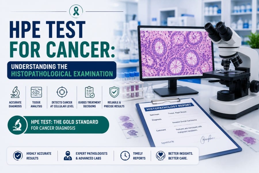
HPE Test for Cancer: Understanding the Histopathological Examination

The HPE test for cancer is a definitive method for confirming malignancy, grading disease, and guiding many decisions. While digital pathology platforms and laboratory informatics evolve, histopathological examination stands to retain its authority in tissue-based examinations. Because cellular architecture still tells the most reliable story.
For professionals working across surgical pathology, oncopathology, or laboratory management, clarity around the HPE test, reporting, and operational issues matters. The discussion below addresses the science, the lab process, and the practical considerations shaping HPE testing across modern pathology labs.
What is the HPE Test for Cancer
The HPE test full form in medical parlance is Histopathological Examination. The test involves microscopic evaluation of tissue sections processed through fixation, embedding, sectioning, and staining. The purpose extends beyond the detection of malignant cells. Tumour margins, invasion depth, lymphovascular spread, and precursor lesions all come into focus under the microscope.
In oncology procedures, the HPE test functions as the final diagnostic step after imaging and tissue acquisition. Cytology and radiology suggest probability. Histopathology confirms the disease.
Biopsy HPE test: from tissue to slide
A biopsy HPE test begins long before slides reach a reporting pathologist. Tissue handling at each stage affects diagnostic fidelity.
Stages in Biopsy HPE Processing
- Specimen receipt with site verification and clinical notes.
- Fixation using buffered formalin to preserve morphology.
- Gross examination and sampling by trained personnel.
- Tissue processing and paraffin embedding.
- Microtomy with section thickness control.
- Staining, commonly with hematoxylin and eosin.
- Optional immunohistochemistry based on differential diagnosis
Each stage introduces variables. Fixation delay alters nuclear detail. Section thickness affects interpretation. Staining consistency shapes pattern recognition.
Therefore, laboratory standardisation is imperative.
HPE Test for Cancer Across Tumour Types
The HPE test for cancer applies across solid malignancies, e.g., Breast, gastrointestinal, lung, gynecologic, genitourinary, and soft tissue tumours.
Breast cancer
The HPE test for breast cancer determines histologic subtype, tumour grade, lymph node status, and margin involvement. These findings influence surgical decisions and adjuvant therapy planning. Hormone receptor studies and HER2 testing build upon the HPE diagnosis.
Breast specimens also illustrate the procedural complexity. Lumpectomy samples demand precise orientation. Sentinel lymph nodes require serial sectioning. The accuracy of reporting depends on structured practices.
Pathology Report Format
For clinicians, the pathology report carries more weight than raw microscopic impressions. A consistent pathology report format supports clinical decision-making and interdisciplinary communication.
Components in a Report
- Patient identifiers and specimen details.
- Clinical history relevant to interpretation.
- Gross description summarising specimen features.
- Microscopic findings with diagnostic terminology.
- Final diagnosis using standard classifications.
- Ancillary study results are performed.
- Comments addressing margins or prognostic features.
Structured reporting reduces ambiguity. Synoptic templates align pathology language with oncology protocols.
Digital reporting platforms like Flabs now integrate these elements directly into laboratory information systems (LISs).
Turnaround Time
In the HPE test for cancer, turnaround time is critical in two ways: as a performance metric for pathology labs and for faster diagnosis. Yet speed cannot compromise interpretive quality. Routine biopsy HPE reports range from 2-5 working days. Complex resections or cases requiring immunohistochemistry extend timelines.
Further, specimen volume, staffing expertise, lab equipment, and digital workflow integration also impact the early availability of results. Transparent communication with clinicians around expected reporting windows maintains trust and reduces follow-up queries.
Lab Equipment and Infrastructure
- Automated tissue processors.
- Paraffin embedding stations.
- Rotary microtomes with blade stability.
- Automated stainers.
- Slide scanners for digital pathology.
- Laboratory information systems integrated with reporting.
Operational Realities
Pathology labs in India operate within a diverse ecosystem. Large reference laboratories coexist with hospital-based departments and independent diagnostic centres. Accreditation standards, workload distribution, and resource availability vary widely among them.
For example, urban centres increasingly adopt digital pathology, AI-assisted screening, and centralised reporting models. Semi-urban and rural labs prioritise turnaround control and cost efficiency.
Fortunately, laboratory software platforms now bridge geographic gaps by enabling remote reporting, workload balancing, and quality audits.
Parameters in routine oncology practice
| Parameter | Relevance |
|---|---|
| Tissue fixation quality | Preserves cellular morphology |
| Section thickness | Influences nuclear and architectural detail |
| Staining consistency | Enables pattern recognition |
| Report structure | Supports clinical interpretation |
| Turnaround time | Affects treatment planning |
| Equipment calibration | Ensures reproducibility |
Final Perspective
The HPE test for cancer is grounded in morphological science while adapting to digital transformation. Pathologists interpret disease through pattern, context, and clinical correlation. Laboratory leaders manage workflows, equipment, and reporting systems that support diagnostic integrity.
For Medical SaaS companies serving pathology teams, success depends on respecting that balance. Technology must align with histopathology practice rather than disrupt it. When software strengthens reporting clarity, optimises turnaround, and integrates seamlessly into laboratory routines, adoption follows naturally.
And Flabs LIS software achieve that effortlessly.
Start Your 5-Day Free Trial of Flabs Pathology Software Today.
Get Started at ₹1!
Try Flabs for a full month for just ₹1.
Follow us on