Liver cirrhosis pathology is classified into four main stages for diagnosis and treatment. It is caused by chronic viral hepatitis, alcohol abuse, and non-alcoholic fatty liver disease (NAFLD). However, with the advancement of treatments for viral hepatitis, the proportion of cirrhosis cases attributable to alcohol abuse and NAFLD has been increasing.
The condition places a large diagnostic load on pathology laboratories. Among chronic hepatic disorders, cirrhosis represents the final structural transformation of long-standing liver injury.
As a pathologist or clinician, you will evaluate biopsies, correlate clinical findings, and communicate disease severity with clarity. There’s a need for accurate interpretation of liver cirrhosis pathology to support staging, prognosis, and treatment planning.
Liver Cirrhosis Pathology: Stages and Features
You begin with a clear liver cirrhosis definition: a chronic liver condition characterised by diffuse fibrosis, regenerative nodules, and altered vascular architecture. Persistent injury from viral infection, alcohol exposure, metabolic disease, or autoimmune damage drives progressive fibrosis. Over time, fibrosis divides hepatic tissue into nodules that disrupt normal blood flow.
Within liver cirrhosis pathology, three structural elements dominate microscopic evaluation.
- Diffuse fibrotic septa bridging portal tracts and central veins.
- Regenerative hepatocellular nodules.
- Distorted sinusoidal and vascular architecture.
- Gross examination also reveals major structural changes. In advanced disease, the liver appears shrunken with nodular external contours. Pathologists describe cirrhosis liver gross pathology as a firm organ with uneven surfaces and fibrous bands separating nodules of varying size.
From a diagnostic perspective, pathology findings must align with patient presentation. Clinical teams evaluate liver cirrhosis symptoms, laboratory abnormalities, and imaging results alongside biopsy interpretation.
Structural Changes Inside the Cirrhotic Liver
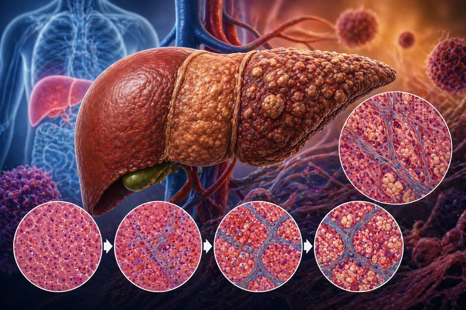
When you evaluate hepatic tissue under microscopy, architectural collapse becomes immediately visible. In a healthy liver, hepatocytes form plates radiating from central veins. Cirrhosis disrupts that arrangement through fibrosis and nodule formation.
Major microscopic findings in liver cirrhosis pathology include the following.
Fibrous Septa Formation: Activated hepatic stellate cells deposit collagen within the space of Disse. Fibrous bands bridge vascular structures and divide the liver into nodules.
Regenerative Nodules: Surviving hepatocytes proliferate in response to injury. These clusters form spherical nodules surrounded by fibrotic tissue.
Vascular Distortion: Portal blood flow bypasses hepatocytes through shunts. Portal hypertension emerges as resistance rises within distorted sinusoids.
Inflammatory Infiltration: Chronic injury leads to lymphocyte and macrophage infiltration in portal regions.
Hepatocyte Degeneration: Ballooning, steatosis, and apoptosis may appear depending on the underlying cause.
Recognition of these features forms the diagnostic core of liver cirrhosis pathology during biopsy interpretation.
Stages of Cirrhosis Progression
Cirrhosis develops gradually over many years. Pathology staging allows clinicians to evaluate fibrosis progression and disease severity. Fibrosis staging systems such as METAVIR and Ishak classify structural changes observed in liver biopsy samples.
| Stage | Pathological Features | Clinical Implications |
|---|---|---|
| Early fibrosis | Portal fibrosis without septa | Minimal symptoms; early liver cirrhosis test findings |
| Fibrosis with septa | Fibrous bridges between portal tracts | Increasing portal pressure |
| Advanced fibrosis | Extensive septa and architectural distortion | Early complications of liver cirrhosis |
| Cirrhosis | Diffuse nodules with fibrotic bands | Portal hypertension and hepatic dysfunction |
During staging assessment, liver cirrhosis pathology focuses on septal thickness, nodule size, and vascular distortion. Accurate staging shapes prognosis and long-term management.
Etiological Patterns in Cirrhosis
Cirrhosis develops from multiple chronic injuries. As a pathologist, you have to interpret morphological patterns that point toward specific etiologies.
Common liver cirrhosis causes include the following.
- Chronic viral hepatitis (HBV, HCV).
- Alcohol-related liver disease.
- Non-alcoholic fatty liver disease.
- Autoimmune hepatitis.
- Metabolic disorders such as hemochromatosis and Wilson's disease.
- Cardiovascular diseases, congestive heart failure and chronic ischemia,
Among these, alcohol exposure produces distinctive morphological changes. Alcoholic liver cirrhosis pathology frequently shows steatosis, Mallory–Denk bodies, neutrophilic infiltration, and perisinusoidal fibrosis.
Recognition of these patterns enhances the interpretation of liver cirrhosis pathology, especially when the clinical history is incomplete.
Diagnostic Evaluation in Clinical Practice
Cirrhosis diagnosis rarely relies on histology alone. Physicians combine pathology with imaging, laboratory markers, and functional scoring systems.
- Liver biopsy with histopathological evaluation.
- Serum liver function tests.
- Elastography or FibroScan.
- CT or MRI imaging.
- Endoscopic screening for varices.
Within diagnostic operations, liver cirrhosis pathology confirms architectural distortion and fibrosis stage. Histology also differentiates cirrhosis from severe fibrosis without nodular regeneration.
Clinical classification also recognises several types of liver cirrhosis.
- Alcoholic cirrhosis.
- Post-necrotic cirrhosis.
- Biliary cirrhosis.
- Metabolic cirrhosis.
Each type presents subtle histological variations.
Clinical Manifestations and Systemic Effects
Cirrhosis produces a wide spectrum of clinical manifestations. Early disease may remain silent, while advanced disease leads to multi-system complications.
Common signs and symptoms of liver cirrhosis include the following.
- Fatigue and weakness.
- Ascites.
- Jaundice.
- Spider angiomas.
- Splenomegaly.
- Gastrointestinal bleeding.
In pathology correlation meetings, you match biopsy findings with patient complaints. Advanced liver cirrhosis pathology correlates strongly with portal hypertension and hepatic insufficiency.
When disease progresses further, clinicians observe end-stage liver cirrhosis symptoms, viz., hepatic encephalopathy, severe ascites, coagulopathy, and renal dysfunction.
Major Complications Associated With Cirrhosis
Chronic structural damage within the liver leads to several life-threatening outcomes.
Major complications of liver cirrhosis include Portal hypertension, Esophageal varices, Gastrointestinal bleeding, Ascites, Hepatorenal syndrome, Hepatocellular carcinoma, and hepatic encephalopathy.
Pathology has a major role in tumour surveillance. Nodular cirrhotic tissue creates a high-risk environment for malignant transformation. Careful evaluation of liver cirrhosis pathology ensures early detection of dysplastic nodules or carcinoma.
Therapy and Lifestyle Measures
Strategies of treatment in liver cirrhosis pathology depend on disease stage and aetiology. The whole clinical management focuses on slowing progression and controlling complications. Currently, liver cirrhosis treatment approaches include the following.
- Antiviral therapy for hepatitis infections.
- Alcohol abstinence programs.
- Immunosuppressive therapy for autoimmune disease.
- Management of portal hypertension.
- Liver transplantation for advanced disease.
Dietary regulation can also help with supportive care. A balanced liver cirrhosis diet generally includes reduced sodium intake, adequate protein, and avoidance of alcohol. Nutritional planning addresses muscle wasting and metabolic imbalance.
In India, tertiary liver centres provide advanced treatment options. Patients with severe disease may require referral to the best hospital for liver cirrhosis in India for transplant evaluation.
Throughout disease management, pathological findings continue to guide treatment decisions. Repeated evaluation of liver cirrhosis pathology assists clinicians in monitoring fibrosis progression and therapeutic response.
Final Perspective
For pathologists and clinicians, cirrhosis represents a complex structural and functional transformation of hepatic tissue. Microscopic interpretation remains central to diagnosis, staging, and etiological identification.
By carefully evaluating liver cirrhosis pathology, you detect fibrosis patterns, regenerative nodules, and vascular distortion that define cirrhosis. These observations guide clinical teams toward appropriate treatment strategies and long-term monitoring.
As chronic liver disease rates continue to rise worldwide, precise interpretation of liver cirrhosis pathology is essential in pathology practice and patient care.
Related read - Liver Function Test Report Format – Free PDF Sample
Get Started at ₹1!
Try Flabs for a full month for just ₹1.
Follow us on