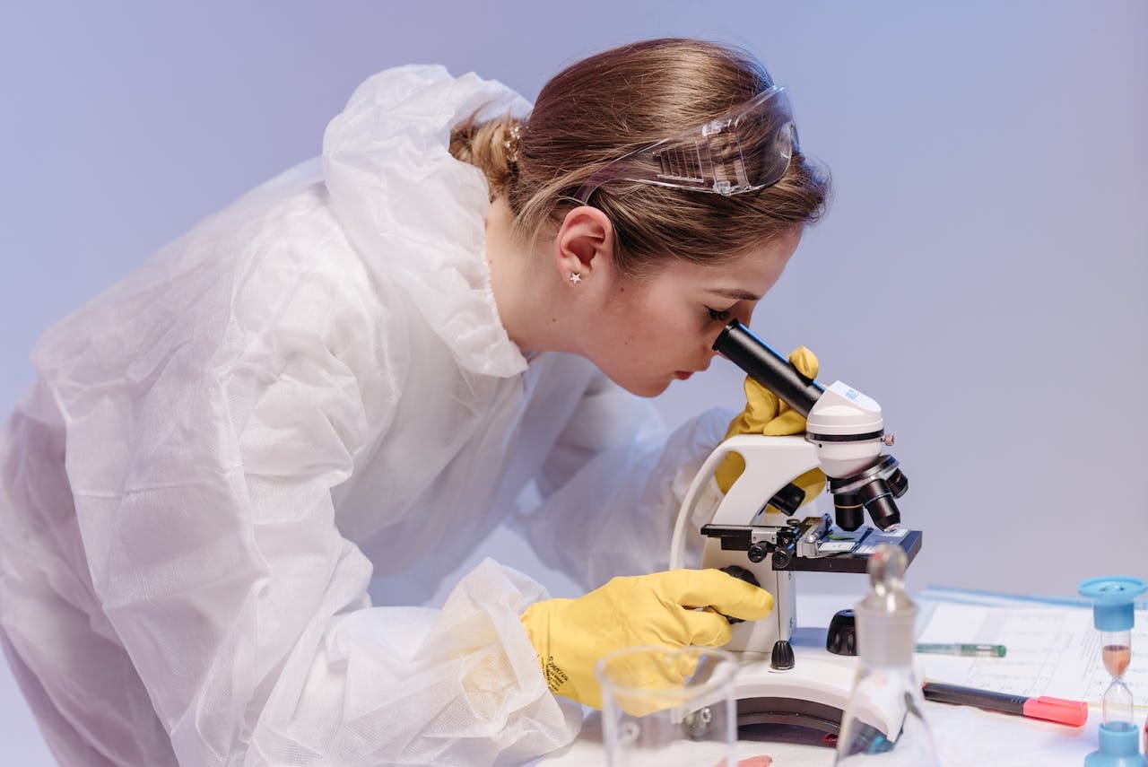
In histopathology, special stains go beyond routine hematoxylin and eosin (H&E) staining, giving visibility into tissue structures and pathologic processes. These specialised techniques use dyes or chemicals tailored to specific tissue components, revealing details that routine stains may miss.
By highlighting features like connective tissue, microorganisms, or pigments, the list of special stains in histopathology helps pathologists identify diseases, assess tissue integrity, and uncover cellular abnormalities.
Essential in both diagnosis and research, they improve the visualisation of otherwise transparent sections, allowing precise evaluation of morphology and specific cell types. Let’s explore the special stains in a histopathology laboratory and their indispensable role in advancing histological examinations.
Special Stains in Histopathology and Importance
- Special stains used in histopathology labs have specific purposes in tissue studies. Some of their main uses are that they:
- Enhance tissue details for better microscopic analysis.
- Identify specific cell structures and tissue components.
- Detect pathogens such as bacteria, fungi, and viruses.
- Highlight abnormal deposits like amyloid and iron.
- Distinguish between different types of cancer.
- Aid in diagnosing conditions not visible with routine stains.
- Improve visualisation of connective tissue, muscle fibers, and nerve cells.
- Assist in evaluating disease progression and treatment responses.
Special Stains in Histopathology and their Uses
| Stain Name | Primary Use | Color Results |
|---|---|---|
| Giemsa Stain | Multiple tissue components identification | Mast cells/Cartilage/Mucin/Amyloid: Purple Background: Blue |
| Wright's Stain | Peripheral blood smears | Similar to Giemsa |
| Hematoxylin and Eosin (H&E) | Routine morphological structure identification | Nuclei: Blue-purple Cytoplasm/Muscle: Pink |
| Movat's Pentachrome | Fibrin, mucin, and collagen visualisation | Nuclei/Elastic fibres: Black Fibrin: Intense red Muscle: Red Collagen/Reticular fibers: Yellow Ground substance mucin: Blue |
| Picrosirius Red | Collagen visualisation | Collagen: Vibrant red/Orange-red Other elements: Yellow |
| Mason's Trichrome | Connective tissue fibres | Collagen: Blue Nuclei: Blue-black Cytoplasm/Keratin/Muscle: Pink to red |
| AZAN Trichrome | Collagen fibers | Nuclei/Erythrocytes: Red Muscle: Orange Collagen/Reticulum: Dark blue |
| Combined Massons Elastin | Connective tissue fibres | Elastic fibres/Nuclei: Black Collagen/Mucin: Green Keratin/Cytoplasm/Muscle: Red |
| Oil Red O | Lipid morphology (frozen sections) | Lipids: Orange to brilliant red Nuclei: Blue to purple |
| Cresyl Echt Violet (CEV) | Nissl substances and nuclei | Nissl substances/Nuclei: Purple Background: Clear |
| Luxol Fast Blue (LFB) | Myelin visualisation | Myelin: Greenish blue Cells: Pink to violet |
| Safranin O | Cartilage, mucins and mast cells | Cartilage/Mucin: Orange to red Nuclei: Black Background: Green |
| PAS Alcian Blue | Glycogen and goblet cells | Glycogen: Rose to red Mucosubstances: Blue |
| PAS & Hematoxylin | Glycogen and goblet cells | Glycogen: Rose to red Nuclei: Blue-violet |
| Mucicarmine | Epithelial mucins | Mucins: Specific staining |
| Von Kossa | Mineralisation | Calcium salts: Black Nuclei: Red/Dark green Cytoplasm: Pink/Green |
| Prussian Blue | Iron deposits | Hemosiderin (iron): Blue Nuclei/Hemofuchsin: Bright red Background: Pink |
| Fontana-Masson | Melanin visualisation | Melanin granules: Black |
| Melanin Bleach | Removal of melanin for cellular morphology | Removes melanin pigmentation |
| Jones Methenamine Silver | Basement membranes | Basement membranes: Black Nuclei: Blue Cytoplasm/Background: Pink |
| Toluidine Blue | Mast cells | Mast cells: Violet to reddish-purple Background: Blue |
| Phloxine/Tartrazine/Alcian Blue | Paneth Cells | Mature cells: Bright red Immature cells: Golden yellow Goblet cells: Pale-light green |
| Chromic Acid Schiff (CAS) | Fungi visualisation | Fungus: Brown exterior, reddish pink interior |
| Ziehl-Neelsen Stain (ZN) | Acid-fast organisms | Acid-fast organisms: Bright pink Background: Methylene blue |
Special Stains in Histopathology Classification
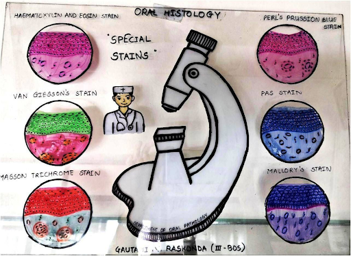
- Basic Staining: Hematoxylin and Eosin (H&E) and Movat's Pentachrome
- Connective Tissue: Picrosirius Red, Mason's Trichrome, AZAN Trichrome, Combined Massons Elastin, Verhoff's Elastic Stain, Reticulin Stain
- General Purpose: Giemsa Stain, Wright's Stain
- Lipid: Oil Red O
- Neural: Cresyl Echt Violet (CEV), Luxol Fast Blue (LFB)
- Glycogen & Mucins: Safrananin O, PAS Alcian Blue, PAS & Hematoxylin, Colloidal iron ("AMP"), Mucicarmine
- Minerals & Pigments: Von Kossa, Prussian Blue, Fontana-Masson, Melanin Bleach
- Basement Membranes: Jones Methenamine Silver,
- Mast Cells: Toluidine Blue
- Paneth Cells: Phloxine/Tartrazine/Alcian Blue
- Fungus and Bacteria: Chromic Acid Schiff (CAS), Ziehl-Neelsen Stain (ZN), Brown and Hopps
Conclusion
In addition to their diagnostic applications, special stains in histopathology help monitor treatment effectiveness. By highlighting cellular changes over time, they help assess how tissues respond to therapies, particularly in chronic conditions like fibrosis or cancer. This dynamic application provides clinicians with ongoing insights, enabling more personalised treatment adjustments.
Suggested : Types of Fixative in Histopathology
Get Started at ₹1!
Try Flabs for a full month for just ₹1.
Follow us on