The spinal cord is one of the most structurally organised parts of your nervous system. Measuring about 45 cm in an adult, it serves as the main conduit for motor and sensory signals between the brain and the rest of the body. But what does it actually look like under a microscope?
Spinal cord histology gives you a detailed, tissue-level view of this structure. It is essential for diagnosing neurological diseases, traumatic injuries, and degenerative conditions. As a pathologist or healthcare professional, the ability to read and interpret histological sections of the spinal cord is a skill that directly shapes diagnostic outcomes.
Spinal Cord Histology: How the Tissue Is Organised
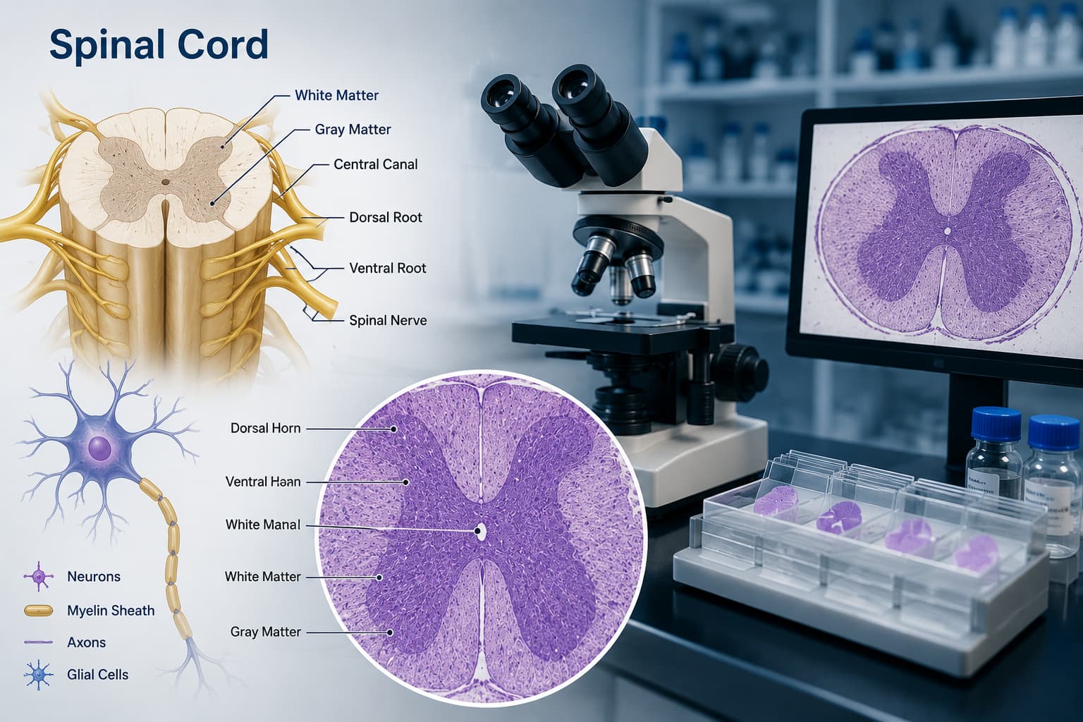
At the centre of every spinal cord cross-section, you see a butterfly- or H-shaped region of grey matter surrounded by white matter. This is the first and most recognisable landmark in spinal cord histology.
The grey matter contains neuronal cell bodies, dendrites, unmyelinated axons, and glial cells. It is divided into three horns: the dorsal (posterior) horn, the ventral (anterior) horn, and the lateral horn (present mainly in thoracic and upper lumbar segments).
The white matter surrounds the grey matter and is composed predominantly of myelinated axons organised into funiculi— the posterior, lateral, and anterior funiculi. White matter in spinal cord histology appears lighter on standard H&E staining because myelin washes out during processing.
The Grey Matter in Detail
The grey matter in spinal cord histology is further organised using Rexed's laminae. It is a system of ten cytoarchitectural layers (I–X) based on neuronal size, density, and arrangement.
Laminae I–VI: Located in the dorsal horn. Lamina I processes nociceptive input; lamina II (substantia gelatinosa) is notable for its small, densely packed neurons.
Laminae VII–IX: Located in the ventral horn. Lamina IX contains the large alpha motor neurons responsible for voluntary movement.
Lamina X: Surrounds the central canal and contains neurons involved in visceral sensation.
In the ventral horn, the alpha motor neurons are large multipolar cells with prominent Nissl substance (rough ER), a large nucleus with a distinct nucleolus, and multiple dendrites. These features are main identifiers in any spinal cord cross-section in histology.
Regional Differences: Cervical, Thoracic, and Lumbar Sections
Spinal cord sections vary significantly across regions. Recognising these differences is essential for accurate diagnosis.
| Feature | Cervical | Thoracic | Lumbar/Sacral |
|---|---|---|---|
| Overall Size | Largest cross-sectional area | Smallest cross-sectional area | Large, especially lumbar enlargement |
| Gray Matter | Large ventral horns (arm muscles) | Small ventral horn; lateral horn present | Massive ventral horns (leg muscles) |
| White Matter | Proportionally very large | Relatively large (long tracts) | Proportionally smaller |
| Lateral Horn | Absent | Present (T1–L2, sympathetic neurons) | Present at S2–S4 (parasympathetic) |
| Distinguishing Feature | Large motor neurons; medial & lateral groups | Clarke's nucleus visible at T1–L2 | Prominent alpha motor neurons |
Cervical spinal cord histology stands out because of its large grey matter relative to white matter, especially in the cervical enlargements (C5–T1), where neurons supplying the upper limb musculature are concentrated.
Thoracic spinal cord histology is characterised by its small overall size, the presence of the lateral horn (housing preganglionic sympathetic neurons), and Clarke's nucleus, a well-defined cluster of large neurons at the base of the dorsal horn visible from T1 to L2.
What You See in a Stained Cross Section
When you look at a spinal cord cross-section in histology, the staining method you use will determine what structures stand out.
- H&E (Hematoxylin & Eosin): Grey matter stains darker (more cellular), white matter appears lighter. Neurons are easily identified by their large soma and purple Nissl bodies.
- Luxol Fast Blue (LFB): The go-to stain for white matter in spinal cord histology. Myelin stains bright blue, making demyelination unmistakably visible.
- Bielschowsky Silver Stain: Highlights axons; useful for identifying axonal loss in degenerative diseases.
- Immunohistochemistry (GFAP, NeuN, MBP): GFAP marks astrocytes, NeuN marks neuronal nuclei, and MBP (Myelin Basic Protein) marks myelinated fibres.
Spinal cord histology labelled sections in atlases are excellent references when you are learning to orient yourself on a new section. Such as those in Netter's or the Allen Spinal Cord Atlas.
Histological Features of the Spinal Cord in Disease
Knowing the normal histological features of spinal cord tissue is what allows you to spot pathology. Here are some conditions and their histological appearances.
Multiple Sclerosis (MS): Demyelinating plaques in the white matter; LFB shows loss of blue staining. Reactive gliosis with GFAP-positive astrocytes.
Amyotrophic Lateral Sclerosis (ALS): Loss of large motor neurons in the ventral horn; degeneration of the corticospinal tracts (lateral funiculi).
Syringomyelia: Abnormal cavity within the cord, usually in the central canal region; surrounded by reactive gliosis.
Spinal cord infarction: Ischemic necrosis with ghost neurons and macrophage infiltration.
Tabes Dorsalis (neurosyphilis): Demyelination and degeneration of the posterior columns.
The histological features of spinal cord tissue in each of these conditions follow distinct patterns. Once you identify the location (white vs grey matter) and nature (demyelination, neuronal loss, gliosis) of the change, you can significantly narrow your differential.
Why Spinal Cord Histology Matters for Labs
Pathology labs in India are seeing a rising volume of neurological biopsies and autopsy specimens, particularly with the increasing burden of stroke, MS, and motor neuron disease. Neuropathology, once a niche subspecialty, is becoming an area where general pathologists need a solid foundation.
Spinal cord histology is a core part of that foundation. Whether you are reviewing a formalin-fixed autopsy section or a surgical specimen from a cord tumour, the ability to identify normal architecture and spot deviations from it is what makes your report clinically actionable.
Labs that invest in upskilling their teams in neuropathology are better positioned to serve neurology and neurosurgery departments effectively.
A Quick Reference: Spinal Cord Histology Landmarks
Central canal: Small, ependymal-lined canal at the centre of the grey matter
Anterior grey commissure: Grey matter connecting the two anterior horns across the midline
Ventral horn: Contains large multipolar alpha motor neurons
Dorsal horn: Contains substantia gelatinosa (lamina II) with small dense neurons
Lateral horn: Present in thoracic/upper lumbar; sympathetic preganglionic neurons.
Anterior funiculus: White matter anterior to the anterior horn.
Lateral funiculus: White matter lateral to the grey matter; contains corticospinal tracts.
Posterior funiculus: White matter posterior to the dorsal horn; contains sensory tracts (fasciculus gracilis and cuneatus).
Run Your Lab Smarter with AI-Powered LIS
Accurate interpretation of spinal cord histology requires not just expertise, but also efficient operations and reliable reporting. In high-volume pathology labs, even small delays or inconsistencies can impact clinical decisions.
Flabs AI-Powered Pathology Software is designed to support labs handling complex cases, including neurological specimens, by streamlining reporting and enhancing diagnostic precision.
From structured reporting for histopathology to intelligent insights that assist in identifying patterns, Flabs LIS helps pathologists focus more on interpretation and less on manual processes.
✔ Standardised Histopathology Reporting.
✔ Faster Turnaround Time for Critical Cases.
✔ AI-Assisted Insights for Better Accuracy.
✔ Seamless Report Sharing with Clinicians.
✔ Cloud-Based Access for Anytime Review.
Whether you're reviewing spinal cord sections or managing a growing diagnostic workload, Flabs helps your lab stay efficient, accurate, and future-ready.
Start your 5-day free trial and experience smarter pathology operations today.
Get Started at ₹1!
Try Flabs for a full month for just ₹1.
Follow us on