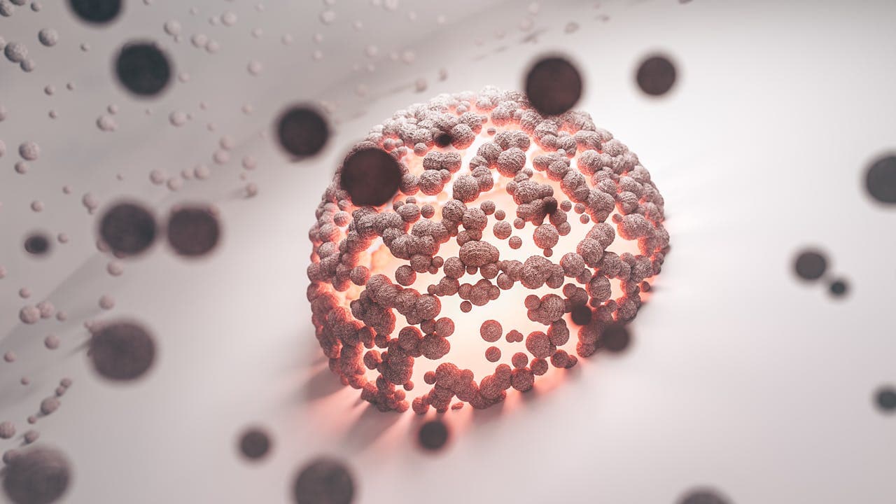
Giant cells are multinucleated cells that arise from the fusion of monocytes or macrophages. They are present in various pathological and physiological contexts. While some types are normal in specific tissues, their presence often signals underlying diseases. For pathologists and healthcare professionals, knowledge of types of giant cells is irreplaceable. They can diagnose and manage conditions ranging from infections to neoplasms.
What Are Giant Cells?
Giant cells are large, multinucleated cells formed when monocytes or macrophages fuse. It can occur in response to infections, foreign bodies, or autoimmune diseases. The fusion mechanism and resulting cell type depend on the triggering stimulus. In some cases, these cells actively participate in tissue repair. In others, they drive or reflect disease pathology.
There are distinct types of giant cells based on morphology, function, and the conditions they arise. Each type provides unique information about the underlying pathology.
Types of Giant Cells
| Giant Cell Type | Associated Conditions | Key Markers* | Formation Mechanism* |
|---|---|---|---|
| Osteoclast-like Giant Cells (OCLGCs) | Giant cell tumour, aneurysmal bone cyst, osteosarcoma | TRAP+, CD68+, RANK+, cathepsin K+ | Driven by RANKL signalling, chromosomal changes, and increased osteoclast activity |
| Langhans Giant Cells | Tuberculosis, granulomatous inflammation | CD68+, DC-STAMP+, E-cadherin+ | Fusion promoted by IFN-γ, CD40 signalling, and intercellular adhesion molecules |
| Foreign Body Giant Cells (FBGCs) | Foreign body giant cell reactions | CD68+, DC-STAMP+, CD206+ | Triggered by IL-4/IL-13; adhesion to foreign material via integrins and E-cadherin |
| Touton Giant Cells | Xanthogranulomas | CD68+, lysozyme+, lipid-filled cytoplasm | Derived from macrophages responding to IL-6, it is often involved in lipid removal |
| Cherubism-associated Giant Cells | Cherubism | TRAP+, V-ATPase+, cathepsin K+ | Mutation in SH3BP2 enhances NFAT signalling, promoting osteoclast-like cell formation |
| Brown Tumor-associated Giant Cells | Hyperparathyroidism | TRAP+, CD68+, RANK+ | Increased RANKL expression due to parathyroid hormone promotes osteoclastogenesis |
| Giant Cell Arteritis-associated Giant Cells | Giant cell arteritis | CD68+, iNOS+, MMP-9 | Driven by IFN-γ and TNF-α, leading to macrophage fusion and vascular tissue damage |
| Sarcoidosis-associated Giant Cells | Sarcoidosis | Arginase+, CD206+, CD44+ | IL-4/IL-13 stimulates macrophage fusion through CD44-mediated interactions |
*Key Markers: These markers help identify the giant cell type and understand its function.
**Formation Mechanisms: Provide insights into how these cells develop and their role in disease processes.
1. Osteoclasts
Source - Research Gate
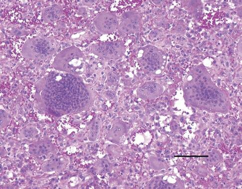
Osteoclasts are specialised types of giant cells. They are essential for bone resorption and remodelling. These cells form through the fusion of preosteoclasts derived from monocytes. Osteoclasts express specific markers like tartrate-resistant acid phosphatase (TRAP) and RANKL. They are commonly found on bone surfaces where resorption occurs. Their primary function is to break down the bone matrix and minerals, enabling bone remodelling and calcium release.
Osteoclasts are involved in normal physiology but can also contribute to diseases. Conditions like osteoporosis, osteosarcoma, and giant cell tumours of bone often feature osteoclast-like activity.
2. Foreign Body Giant Cells (FBGCs)
Source - Wikipedia
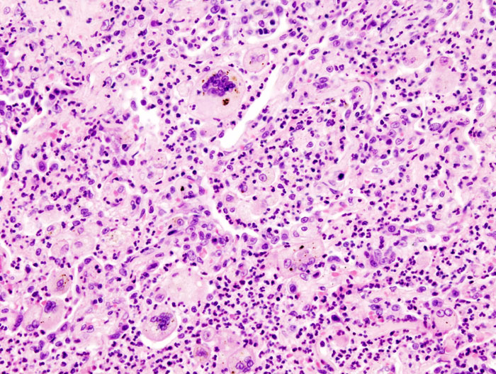
Foreign body giant cells form in response to non-degradable foreign materials. Examples of these types of giant cells include surgical implants, suture material, and other foreign bodies. These cells arise from the fusion of macrophages and are driven by interleukins like IL-4 and IL-13. FBGCs express markers such as CD68 and DC-STAMP.
FBGCs are highly phagocytic but often fail to digest foreign material. Their formation represents the body's attempt to isolate and contain the material. They are commonly seen in granulomas near foreign bodies. Their presence may indicate an unresolved inflammatory response.
3. Langhans Giant Cells
Langhans giant cells are spotted through granulomatous inflammation. These types of giant cells are often seen in infections like tuberculosis and leprosy. They are also present in sarcoidosis and certain fungal infections. Langhans giant cells’ nuclei are arranged in a peripheral, horseshoe-like pattern.
These cells form under cytokines like interferon-γ and tumour necrosis factor-α. They are highly phagocytic and play a role in containing infectious agents. However, these types of giant cells often reflect chronic infections. Langhans giant cells express markers like CD68 and inducible nitric oxide synthase (iNOS).
4. Touton Giant Cells
Touton giant cells are associated with lipid-laden lesions. They are often seen in xanthomas, xanthogranulomas, and other lipid-rich environments. These cells display a unique appearance. Their nuclei form a circular pattern surrounded by foamy cytoplasm.
Touton giant cells are derived from macrophages. Their formation is influenced by interleukins like IL-6 and lipopolysaccharides (LPS). These types of giant cells play a reparative role, removing extracellular lipids.
Roles of Giant Cells in Pathology
Giant cells serve diverse roles in pathology. Their functions depend on their type and the disease context. Some key roles include:
- Bone Resorption: Osteoclasts break down bone in normal remodelling and pathological conditions.
- Foreign Material Containment: FBGCs isolate and attempt to digest non-degradable materials.
- Infection Control: Langhans cells help contain and limit the spread of pathogens in chronic infections.
- Lipid Clearance: Touton cells remove excess lipids from tissues.
Diagnostic Value of Giant Cells
Giant cell pathology is possible because of their presence, morphology, and markers.
- Langhans Giant Cells: Suggest granulomatous diseases like tuberculosis or sarcoidosis.
- Touton Giant Cells: Indicate lipid-related lesions such as xanthogranulomas.
- Osteoclast-like Giant Cells: Found in bone tumours or metabolic bone diseases.
- Foreign Body Giant Cells: Signal the presence of non-degradable foreign material.
Mechanisms of Giant Cell Formation
Giant cell formation involves three main stages: priming, migration, and fusion. Specific cytokines and adhesion molecules regulate each stage. For example, RANKL drives osteoclast formation, while IL-4 promotes FBGC development. Fusion competence requires the expression of fusogens like DC-STAMP and E-cadherin.
Actin-rich projections called fusopods facilitate cell approximation and adhesion. Phospholipids like phosphatidylserine enhance membrane fluidity, enabling fusion. It e tightly regulated to ensure proper giant cell formation.
Challenges in Giant Cell Research
The molecular pathways of giant cells and their roles in diseases are not fully defined. Histological similarities between types can complicate diagnosis. Moreover, there is a lack of targeted therapies for some conditions related to them. So, treatment options are few and far between.
There is a need for research to identify markers for each giant cell type. Currently, treating diseases involving giant cells is said to rely on targeted therapies, and they are yet to be developed.
Conclusion
Giant cells are a fascinating and complex component of pathology. Yet, exploring their characteristics and functions is indispensable. Advances in molecular diagnostics and targeted therapies are promising to address giant cell-associated diseases. For pathologists and healthcare professionals, a more profound knowledge on all types of giant cells opens new doors.
Get Started at ₹1!
Try Flabs for a full month for just ₹1.
Follow us on