Tissue staining is one of the most sensitive steps in histopathology labs. It refines slide preparation, directly impacting the clarity of diagnostic interpretation. While manual processes are error-prone, automated stainers have become essential for efficient staining solutions in labs. Designed to eliminate variability and minimise human intervention, automated slide stainers are reshaping how labs approach staining procedures.
Let's explore how automated slide stainers function, their advantages, and why more laboratories in India and globally are integrating them into their workflows.
What Is an Automated Stainer?
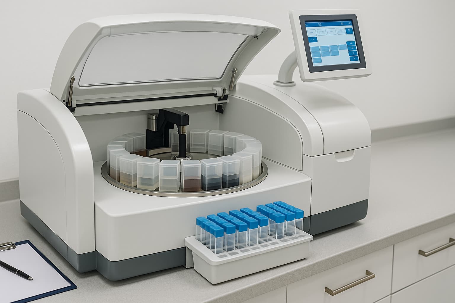
An automated stainer, or automatic slide stainer, is a pathology lab equipment used to apply staining reagents to histology slides in a fully automated manner. These machines perform repetitive and time-sensitive staining tasks with accuracy, controlling each stage, e.g., reagent dispensing, incubation timing and rinsing. In pathology labs, such automation ensures repeatable results.
Working Principle of an Automated Slide Stainer
The automated stainer's working principle involves a programmable sequence of steps.
| Step | Description |
|---|---|
| Slide Loading | Prepared slides are placed in trays or racks designed for the stainer. |
| Reagent Dispensing | The stainer machine accurately dispenses precise volumes of staining reagents. |
| Incubation | Slides are incubated at controlled temperatures for set durations as programmed. |
| Washing and Drying | Excess reagents are rinsed off, and slides are dried or moved to the next step. |
| Unloading | Finished slides are ready for microscopic examination or digital scanning. |
Common Staining Techniques
Automated stainers are designed to support a broad spectrum of staining methods required in histopathology and molecular diagnostics.
1. Hematoxylin and Eosin (H&E) Staining
H&E staining is foundational in pathology. Hematoxylin stains cell nuclei blue-purple, while eosin stains cytoplasm pink. With an automated slide stainer, each step—application, timing, and washing—is performed with machine precision.
2. Immunohistochemistry (IHC)
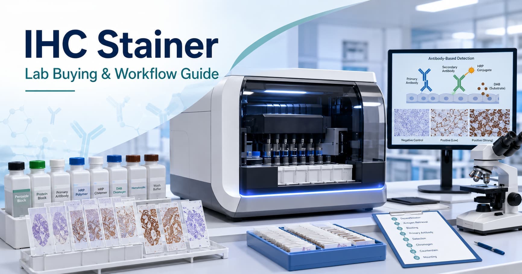
IHC detects specific proteins using antibodies and is indispensable in cancer diagnostics. The automatic slide stainer in histopathology handles incubation with primary and secondary antibodies, chromogen development, and precise temperature control.
3. Special Stains
Special staining methods like Masson’s trichrome, PAS, and silver staining highlight particular tissue structures or pathologies. Automated stainers simplify these complex protocols by automating timing, reagent exposure, and rinse steps to maintain fidelity and reproducibility.
4. Fluorescence In Situ Hybridisation (FISH)
FISH identifies genetic alterations using fluorescent probes. Automated stainers with fluorescence capabilities streamline the hybridisation and wash steps involved in FISH protocols, ensuring accuracy in detecting gene amplifications or chromosomal rearrangements.
Advantages of Using an Automated Slide Stainer
- Automated slide stainers provide consistent and standardised staining, reducing variation seen in manual methods.
- Stainer machines save time by handling multiple slides at once, allowing lab staff to focus on other tasks.
- Automated reagent dispensing controls usage, reduces waste, and prevents contamination. It is imperative when using expensive or limited reagents for special stains and immunohistochemistry.
- Many automated slide stainers from top automated stainer manufacturers connect easily with LIMS and digital imaging systems.
- These systems are scalable, making them suitable for both small labs and large hospitals.
Selecting an Automated Stainer
When choosing an automatic slide stainer, consider the following:
- Volume of slides to be processed daily.
- Types of stains required (H&E, IHC, FISH, special stains).
- Compatibility with digital pathology systems.
- Ease of use for routine operations.
- Maintenance support and service availability.
- Automated stainer price based on feature sets and usage scale.
Integration with Histopathology Workflow
A modern histopathology lab rarely relies on a single instrument. Alongside slide stainers, instruments like tissue processors, embedding centres, cryostats, microtomes, and slide scanners form an integrated system. Efficient staining connects upstream preparation (e.g., tissue sectioning) with downstream analysis (e.g., AI-assisted image review), enabling a seamless workflow.
The synergy between automated devices improves throughput and reduces turnaround time for pathology reports.
Conclusion
Automated stainers not only improve staining efficiency but also support training and skill development. Technicians can focus on interpreting staining results rather than worry about variability. In academic institutions, consistent slides improve learning in histological evaluation.
As digital pathology expands, the precision of automated staining ensures compatibility with AI-based image analysis. For labs planning to scale or adopt advanced diagnostic tools, investing in a reliable automated slide stainer lays a strong foundation for future-ready workflows in histopathology.
Also chekc - Automated Analysers in Modern Pathology Labs
Get Started at ₹1!
Try Flabs for a full month for just ₹1.
Follow us on