Cytopathology examines cells from various body sites to detect abnormalities that may indicate disease. Cytopathology tests are a screening and diagnostic method for numerous conditions, including malignancies. The examination of cellular components, rather than tissue architecture, allows pathologists to identify specific cellular changes.
For healthcare professionals, knowing the applications, limitations, and advancements in cytopathology provides advantages in patient care. This post explores cytopathology tests in depth, examining their methodology, applications in modern clinical practice.
What is Cytopathology?
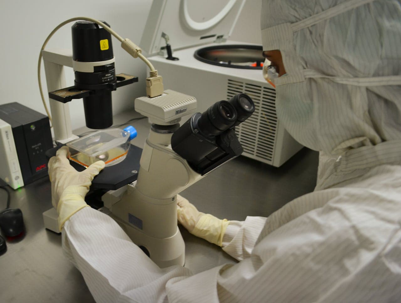
Cytopathology or cytology is the branch of pathology that studies diseases at the cellular level. The cytopathology meaning encompasses the examination of cells from various body fluids and tissues to detect abnormalities in cell structure, organisation, and function.
Unlike other approaches that require larger tissue samples, cytopathology focuses on individual cells or small cell clusters. The specialised field enables pathologists to identify cellular changes associated with inflammatory conditions, infections, benign neoplasms, precancerous lesions, and malignancies.
The practice of cytopathology began in the early 20th century with Dr. George Papanicolaou's development of the Pap smear for cervical cancer screening. Since then, the field has expanded dramatically to encompass numerous specimen types and applications.
Cytopathology vs. Histopathology
The difference between cytopathology and histopathology lies in their fundamental approach to examining a biological material.
| Cytopathology | Histopathology |
|---|---|
| Examines individual cells or small cell clusters | Studies tissue architecture and organisation |
| Requires smaller samples | Requires larger tissue specimens |
| Less invasive collection methods | More invasive collection methods |
| Focuses on cellular characteristics | Examines tissue patterns and relationships |
| Rapid processing (often same-day results) | Longer processing time (typically 1-3 days) |
| Excellent for screening | Gold standard for definitive diagnosis |
Both approaches complement each other in clinical practice. Cytopathology serves as an initial screening tool, while histopathology provides definitive diagnosis when necessary. In many cases, healthcare providers use cytopathology first due to its less invasive nature and faster turnaround time.
Cytopathology Test List
Test Name
Purpose / Sample Site
Pap Test (Papanicolaou Test)
Screening for cervical cell abnormalities
Fine Needle Aspiration (FNA) – Pathologist
Sampling of masses or lumps by a pathologist
Fine Needle Aspiration (FNA) – Clinician
Sampling of lesions by a medical clinician
Urine Cytology
Detecting abnormal cells in the urinary tract
Bronchoalveolar Lavage (BAL)
Examining lung fluid for infections or malignancy
Cerebrospinal Fluid (CSF) Cytology
Analysing spinal fluid for central nervous system diseases
Sputum Cytology
Evaluating lung secretions for cancer cells
Skin Scrape or Discharge
Diagnosing skin lesions or infections
Cyst Fluid Cytology
Assessing cyst contents for malignancy
Brush Biopsy
Collecting cells from internal organs (e.g., lungs, oesophagus)
Body Cavity Fluid Cytology
Studying pleural, peritoneal, or pericardial fluids
Washing Cytology
Rinsing internal organs (e.g., bladder, stomach) to collect cells
Anal Cytology (Anal Pap)
Screening for cell changes in the anal canal
Types of Cytopathology Specimens
Cytology tests rely on collecting cells from various parts of the body, depending on the clinical need. The type of specimen and collection method can vary based on the organ system and diagnostic goals.
Exfoliative Cytology
It involves collecting cells that naturally shed or are lightly scraped from epithelial surfaces. Common uses include:
- Gynecological cytology – such as cervical or vaginal Pap smears
- Respiratory tract samples – sputum and bronchial washings
- Urinary tract samples – voided urine or bladder washings
- Gastrointestinal sources – including biliary brushings or pancreatic secretions
Fine Needle Aspiration (FNA) Cytology
FNA uses a very thin needle (22–25 gauge) to draw cells from a mass or lesion. FNA is minimally invasive, often guided by ultrasound or CT imaging.
It is commonly used to sample:
- Thyroid nodules
- Breast lumps
- Lymph nodes
- Salivary gland lesions
- Lesions in organs like the lung, liver, and pancreas
Body Fluid Cytology
It evaluates cells present in fluid collections. Fluids examined include:
- Pleural fluid – from around the lungs.
- Peritoneal fluid – from the abdominal cavity.
- Pericardial fluid – from around the heart.
- Cerebrospinal fluid (CSF) – from around the brain and spinal cord.
- Synovial fluid – from joints.
Brushing and Washing Cytology
Cells are collected by brushing the inside of organs or by washing cavities with fluid. These methods are used in:
- Bronchial and oesophageal brushings
- Gastric washings
- Ductal lavage of the breast or salivary ducts
Direct Sampling Techniques
These involve collecting surface cells directly using tools such as brushes or spatulas (e.g., for a Pap test). Some techniques include:
- Scraping the epithelial surface (common in cervical cytology).
- Washing a cavity and retrieving the fluid (e.g., bladder washings).
- Brushing internal ducts or hollow organs.
Processing Cytopathology Specimens
After collection, specimens undergo processing before microscopic examination.
1. Conventional Smear Preparation
Cellular material is directly spread onto a glass slide at the time of collection. The smear is either immediately fixed using alcohol-based fixatives or left to air-dry, depending on the staining technique to be used. Common stains include Papanicolaou and Diff-Quik, which help visualise cellular details for cytological evaluation. It is used for rapid assessments.
2. Liquid-Based Cytology (LBC)
In liquid-based cytology, collected cells are placed into a vial containing a preservative solution. The sample undergoes processing to remove excess blood, mucus, and debris, which enhances clarity. The result is a thin, uniform layer of cells on the slide, improving visualisation.
3. Cell Block Preparation
Cell block preparation involves concentrating cellular elements and embedding them in paraffin wax, similar to histology specimen processing. It preserves cell architecture. The cell block technique is particularly valuable when a tissue-like appearance is necessary.
Cytopathology Test for Cancer
Cytopathology is extensively used in cancer detection and management. It enables early detection through procedures like cervical cancer screening via Pap tests, lung cancer screening in high-risk populations using sputum cytology, bladder cancer surveillance through urine cytology, and pancreatic cancer detection with pancreatic juice cytology.
For diagnostic purposes, cytopathology facilitates evaluation of palpable masses through FNA, investigation of pleural and peritoneal effusions, assessment of pulmonary lesions via bronchial washings or brushings, and examination of cerebrospinal fluid for metastatic disease.
Beyond initial diagnosis, cytopathology serves monitoring applications including post-treatment surveillance for recurrence, evaluation of treatment response, and monitoring disease progression in patients with known malignancies.
Cytology vs Biopsy: When to Use Each
| Factor | Cytology | Biopsy |
|---|---|---|
| Invasiveness | Minimally invasive | More invasive |
| Sample size | Small cellular sample | Larger tissue sample |
| Processing time | Rapid (often same-day) | Longer (1–3 days) |
| Cost | Generally lower | Generally higher |
| Diagnostic yield | May have sampling limitations | More comprehensive |
| Architectural context | Limited | Preserved |
| Application | Ideal for screening, initial evaluation | Definitive diagnosis |
Cytology tests often precede biopsies in the diagnostic algorithm. For example, a thyroid nodule might first undergo FNA cytology, with surgical biopsy reserved for inconclusive results or when architectural context becomes necessary for diagnosis.
Interpretation of Test Results
Cytology test results are structured around several components.
First, a specimen adequacy assessment is made to ensure the sample has sufficient cellularity, proper preservation, and the presence of appropriate cell types. It also checks for any interfering elements that could compromise interpretation.
Next, descriptive findings are reported. They detail the cellular composition, observed morphological features, presence of any microorganisms, and relevant background elements.
The diagnostic interpretation section classifies findings into categories such as normal/negative, reactive or inflammatory, atypical or indeterminate, suspicious for malignancy, or positive for malignancy. In some cases, specific disease processes may be identified.
Finally, the report includes recommendations.
Cytopathology reports also facilitate communication between pathologists and clinicians. Hence, standardised reporting systems like The Bethesda System for cervical cytology and The Paris System for urinary cytology are used for reporting.
Advantages and Limitations of Cytopathology Tests
| Advantages | Limitations |
|---|---|
| Less invasive than tissue biopsy | Limited architectural context |
| Rapid turnaround time | Potential sampling errors |
| Cost-effective | Variable sensitivity depending on specimen type |
| High specificity for malignancy | Requires experienced cytopathologists |
| Applicable to multiple body sites | May yield indeterminate results requiring follow-up |
| Allows immediate assessment of sample adequacy | Difficult distinction between certain entities |
| Supports ancillary testing techniques (e.g., IHC, PCR) |
Advanced Techniques in Cytopathology
Immunocytochemistry
Immunocytochemistry uses antibodies to detect specific proteins within cells. It can help identify the type of cells and distinguish between benign and malignant changes. The method can determine where a tumour originated in cases of metastatic cancer.
Molecular Testing
Molecular techniques analyse genetic material in cells. Methods like FISH (Fluorescence In Situ Hybridisation), PCR, next-generation sequencing, and microarray analysis can detect mutations or chromosomal changes.
Digital Cytopathology
With digital cytopathology, high-quality images of samples are captured and shared electronically. It allows for remote consultations and supports tools like computer-assisted screening and AI applications.
Flow Cytometry
Flow cytometry examines cells in a liquid suspension to study their characteristics. It is commonly used to identify blood cancers by checking specific markers on cell surfaces. It also helps analyse DNA content and the cell cycle.
Conclusion
Cytopathology remains essential in modern diagnostics through its minimally invasive approach to disease detection at the cellular level. As technology advances with digital imaging and artificial intelligence integration, cytopathology continues to evolve. For healthcare professionals, mastering cytopathology interpretation improves patient care through efficient and cost-effective disease assessment.
Get Started at ₹1!
Try Flabs for a full month for just ₹1.
Follow us on