Flabs
Decalcification in Histopathology: Why It Matters
Decalcification removes minerals from calcified tissues like bone. In the medical parlance, decalcification in histopathology has a significant bearing. It allows preparation of paraffin sections that preserve microscopic structures needed for diagnosis. Pathologists perform it after fixation and before paraffin processing. Let’s explore the process, outline monitoring methods, and lists suitable reagents. Properly following these steps fixes problems related to tissue preservation and supports better diagnostic outcomes.
The Basic Structure of Bone
Bone tissue consists of cells (aka. osteocytes) embedded in a calcified matrix. The matrix is reinforced by Type 1 collagen fibers. It is well known that Calcium (Ca) provides the rigidity of bone, in the form of hydroxyapatite crystals. These crystals are dissolved during decalcification. It leaves a cohesive tissue structure resembling dense connective tissue.
Two types of mature bone require distinct approaches:
- Cortical Bone: Dense and structured, forming the shafts of long bones.
- Cancellous Bone: Composed of delicate trabeculae. It is found in areas like the epiphyses of long bones and vertebrae.
The balance of cortical and cancellous bone in a specimen determines the time required for bone decalcification and processing.
Fixation of Bone
Proper fixation is laid importance on for preserving the tissue's structural integrity during decalcification. Poor fixation can lead to tissue maceration and suboptimal staining, particularly in bone marrow areas. Buffered formalin is a standard fixative. However, alternatives like Bouin or Davidson’s fixative may be preferred.
Fixation of bone involves:
- Removing soft tissues for better fixative penetration.
- Slicing bone specimens into thin sections using fine-tooth saws to minimize damage.
- Allowing extended fixation times for optimal results.
Decalcifying Agents
Decalcifying agents in histopathology are the chemicals used to remove calcium or calcium compounds from tissues. They fall into three main categories: Strong Acids, Weak Acids, and Chelating agents.
| Category | Agent | Formula | Characteristic |
|---|---|---|---|
| Strong Acids | Nitric Acid | 5% in distilled water | Fast but can damage tissue if overused. |
| Strong Acids | Perenyi’s Fluid | Nitric acid 40 ml, Chromic acid 30 ml, Alcohol 30 ml | Slower than pure nitric acid but still fast. Overuse affects staining. |
| Strong Acids | Hydrochloric Acid | 5-10% in distilled water | Rapid. Must wash formalin off specimens to avoid harmful byproducts. |
| Strong Acids | Von Ebner’s Solution | Sodium chloride 50 ml, Water 42 ml, Hydrochloric acid 8 ml | Quick but can damage tissue if used too long. |
| Weak Acids | Formic Acid | 10% in distilled water | Gentle, preserves tissue and staining quality. |
| Weak Acids | Evans and Krajian | Formic acid 25 ml, Sodium citrate 10 g, Water 75 ml | Buffered with citrate for effective decalcification. |
| Weak Acids | Kristensen | Formic acid 18 ml, Sodium formate 3.5 g, Water 82 ml | Buffered with formate for gentle action. |
| Weak Acids | Gooding and Stewart | Formic acid 5-25 ml, Formaldehyde 5 ml, Water 75 ml | Fixes and decalcifies at the same time. |
| Chelating Agents | Neutral EDTA | EDTA 250 g, Water 1750 ml, Adjust to pH 7.0 | Very gentle but slow. Best for research and molecular techniques. |
Methods of Decalcification in Histopathology
Several methods are employed in histopathology to achieve decalcification. These approaches aim to completely remove calcium salts. As discussed, they must also preserve tissue morphology and yield themselves to staining techniques. Below are the primary methods:
1. Acid Decalcification
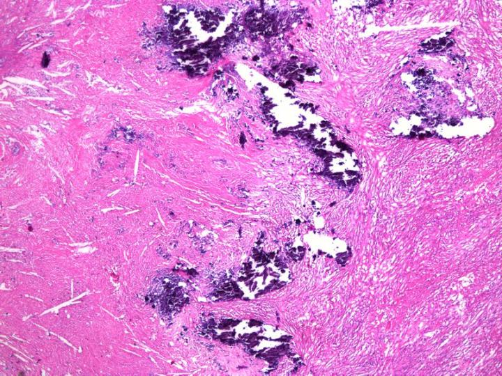
Source - Leica Biosystems
Acid decalcification is the most commonly used method. Combining with neutralisers, various acid solutions are applied. Neutralisers prevent swelling of cells. Common acid decalcifiers include:
- Aqueous Nitric Acid: A rapid agent offering good nuclear staining. But it requires careful monitoring to prevent tissue damage.
- Nitric Acid Formaldehyde: Combines nitric acid and formalin for rapid action and tissue protection from maceration.
- Formic Acid Solution: It is gentler than nitric acid. While it preserves tissue morphology, it decalcifies more slowly.
- Trichloroacetic Acid: Suitable for small biopsies but ineffective for dense or large bony specimens.
2. Ion Exchange Method
This method of decalcification in histopathology uses ammonium salts of sulfonated polystyrene resin. The resin absorbs calcium ions and promotes faster decalcification. It’s particularly effective for research applications requiring detailed morphology.
3. Electrolytic Method
Formic acid or hydrochloric acid serves as the electrolytic medium in the electrolytic method. Calcium ions are drawn to the cathode and enable rapid decalcification. However, heat generated during the process can damage cytological details. So, it is less commonly used.
4. Chelating Agents
Chelating agents (e.g. EDTA) bind calcium ions to form a non-ionised soluble complex. Hence, it is ideal for preserving cytological details in bone marrow biopsies. Although slow, it’s preferred for research and applications relying on molecular integrity.
5. Surface Decalcification
Surface decalcification in histopathology addresses unanticipated calcium deposits in paraffin blocks. The block’s surface is treated with 5% HCl for a brief period. It leads to only superficial decalcification. You can obtain better-quality sections from challenging specimens.
Procedure of Decalcification
- Tissues should be thoroughly fixed before starting decalcification. Proper fixation prevents maceration and ensures good staining.
- Large specimens require the removal of soft tissues for better fixative penetration.
- Slice bone specimens into thin sections using fine-tooth saws to improve fixation and decalcification.
- Select the appropriate decalcifying agent based on the type of tissue and desired outcome.
- Use a sufficient amount of decalcifying solution. Maintain a ratio of solution to tissue volume and replace the solution regularly.
- Monitor the process carefully to avoid incomplete or over-decalcification.
- Apply specific techniques for different scenarios. It might require observation and experience.
- Use surface decalcification for paraffin blocks with unexpected calcium deposits. Refrigerate specimens in decalcifying solution at 4°C to slow down the process if needed.
- After decalcification, wash tissues thoroughly with water.
- For dense or compact bone specimens, use an extended processing schedule. It facilitates proper infiltration of the tissue.
- Apply vacuum during wax infiltration. It improves the quality of processed blocks.
- Avoid over-decalcification.
- Treat with solutions like saturated lithium carbonate or sodium bicarbonate for over-decalcified tissues. They can restore staining quality.
Factors Influencing Decalcification
Decalcification in histopathology is not a self-contained procedure. Several factors determine the outcomes:
Concentration: Higher concentrations act faster. But it may increase tissue damage.
Temperature: Elevated temperatures speed up decalcification. There is also a risk of morphological damage.
Agitation: Gentle agitation improves the process.
Fluid Access: Adequate solution volume and specimen exposure facilitate thorough decalcification.
The End Point of Decalcification in Histopathology
The endpoint of decalcification in histopathology confirms complete calcium removal from tissue samples. X-ray testing is the most precise method. It reveals any remaining calcium clearly. Chemical testing is another reliable approach. It identifies calcium in the solution. Though Physical testing is used, the bending or probing is less reliable. It can also damage the sample.
If bone decalcification needs to be paused, specimens can be rinsed and stored in formalin or refrigerated in the decalcifier.
Points to Take Note
- After decalcification, wash the specimen thoroughly in water to remove residues.
- Over-decalcification can lead to poor nuclear staining and reduced quality.
- Acid-based solutions are used to dissolve calcium salts and soften the bone.
- Zenker’s solution is commonly used for bone marrow specimens to prevent nucleic acid hydrolysis.
- The decalcification process produces carbon dioxide gas as a natural byproduct.
- Bone sections should be cut into 4–5 mm pieces to allow proper penetration of the solution.
- Agitating the decalcifying solution gently helps speed up the process.
- Avoid leaving specimens in the solution for too long, as it can damage tissue morphology.
- Always handle the tissues carefully to preserve their integrity during decalcification.
Closure
Decalcification in histopathology is an established procedure. However, the molecular applications of decalcification go beyond that. Recent advances in single-cell RNA sequencing and spatial transcriptomics have underlined the need for proper decalcification.
These cutting-edge technologies require exceptionally well-preserved tissue architecture and molecular integrity. Expertise in decalcification methods in histopathology is highly recommended for diagnostic and research purposes. It has immense scope in personalised medicine and tissue engineering.
You may also like to read - Special stains used in histopathology and different tests
Get Started at ₹1!
Try Flabs for a full month for just ₹1.
Follow us on