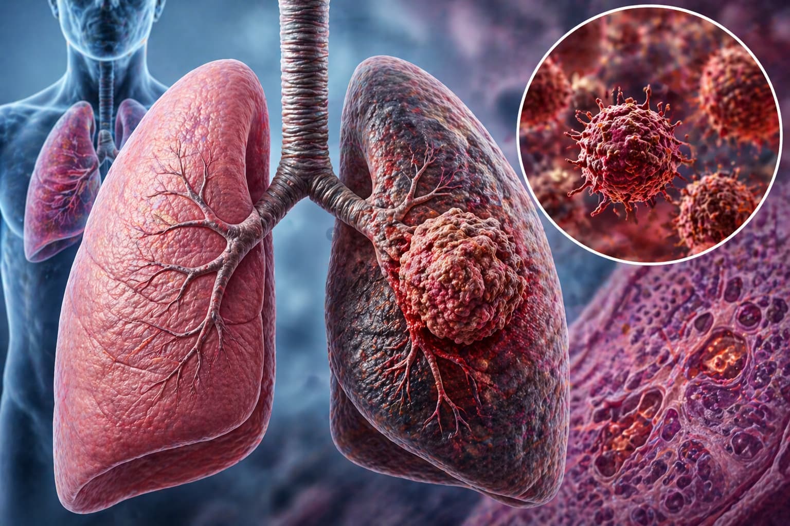
Lung cancer kills more people globally than any other cancer. More than breast, prostate, and colon cancers combined. As a pathologist or healthcare professional, your diagnosis is the starting point for every treatment decision that follows. Before a patient receives a single dose of chemotherapy or steps into a radiation suite, your report defines the road ahead.
So let’s get into it. What you need to know about lung cancer pathology, the types, their defining features, and how accurate classification changes patient outcomes.
What Is Lung Cancer Pathology and Why Classification?
Lung cancer pathology is the microscopic and molecular examination of lung tissue to identify the cancer type, grade, and characteristics that drive treatment planning. A wrong or incomplete classification can mean the wrong drug, the wrong dose, or a missed targetable mutation.
Lung cancer broadly divides into two major groups: Non-Small Cell Lung Cancer (NSCLC) and Small Cell Lung Cancer (SCLC). These two groups behave differently, respond to different therapies, and carry different prognoses.
The Major Types: Pathological Classification of Lung Cancer
There are many types of cancers that affect the lungs. But only two main kinds are considered: Non-Small Cell Lung Cancer (NSCLC) and Small Cell Lung Cancer (SCLC).
Non-Small Cell Lung Cancer (NSCLC)
NSCLC accounts for roughly 85% of all lung cancer cases. It further splits into three primary subtypes.
1. Adenocarcinoma: The most common subtype in non-smokers and women. Adenocarcinoma arises from the glandular epithelial cells—you’ll find it peripherally in the lung. Histologically, it shows acinar, papillary, micropapillary, lepidic, or solid growth patterns.
The lepidic pattern, where tumour cells grow along alveolar walls without destroying the architecture, is specific to this subtype. EGFR, ALK, ROS1, and KRAS mutations are frequent molecular targets here.
2. Squamous Cell Carcinoma (SCC): Strongly linked to smoking. Squamous cell carcinoma originates in the central airways and shows keratinisation, intercellular bridges, and keratin pearl formation under the microscope. IHC markers p40, p63, and CK5/6 are the most reliable for SCC. FGFR1 amplification is a notable molecular finding.
3. Large Cell Carcinoma: A diagnosis of exclusion. When a tumour fails to show features of adenocarcinoma, squamous cell carcinoma, or SCLC, it gets classified as large cell carcinoma. It tends to be aggressive, centrally or peripherally located. It also shows no glandular or squamous differentiation.
Small Cell Lung Cancer Pathology
Small cell lung cancer (SCLC) is the most aggressive form, accounting for about 13–15% of lung cancers. It’s almost exclusively linked to heavy tobacco use.
Under the microscope, SCLC shows:
- Small, oval to fusiform cells.
- Scant cytoplasm.
- Finely granular nuclear chromatin (“salt and pepper” appearance).
- Absent or inconspicuous nucleoli.
- High mitotic rate and extensive necrosis.
SCLC is a neuroendocrine tumour. CD56, synaptophysin, and chromogranin A are positive on IHC. TTF-1 is positive in about 90% of cases.
Because SCLC spreads rapidly, it’s rarely resectable by the time of diagnosis. The TNM system applies. But clinically it is staged as limited (one hemithorax) or extensive (beyond one hemithorax).
Lung Cancer Symptoms and Their Pathological Correlates
Symptoms of lung cancer depend closely on the location of the tumour and type.
Central tumours (SCC, SCLC): Cough, hemoptysis, post-obstructive pneumonia, wheeze.
Peripheral tumours (adenocarcinoma): Often asymptomatic in early stages; pleuritic chest pain in advanced disease.
Pancoast tumours: Shoulder pain, Horner’s syndrome.
Paraneoplastic syndromes: SIADH and Cushing’s syndrome in SCLC, hypercalcemia in SCC.
Stage 1 lung cancer symptoms are largely absent or non-specific, viz., fatigue and mild cough. But it underscores why incidental detection and thorough evaluation of every specimen matters.
Lung Cancer Gross Pathology: What You See Before the Microscope
Gross examination gives you the first clues about tumour type and origin.
| Feature | Adenocarcinoma | Squamous Cell Carcinoma | SCLC |
|---|---|---|---|
| Location | Peripheral | Central (hilar) | Central |
| Gross appearance | Gray-white, often pleural puckering | Gray-white, cavitation possible | Soft, grey, bulky hilar mass |
| Borders | Irregular, spiculated | Irregular | Infiltrative |
| Associated findings | Satellite nodules, pleural effusion | Hilar lymph node involvement | Mediastinal involvement |
| Size at diagnosis | Variable | Variable | Usually large |
Reviewing the lung cancer X-ray alongside gross findings is standard practice. On imaging, peripheral masses with spiculated margins point to adenocarcinoma, while central masses with hilar prominence suggest squamous cell carcinoma or SCLC.
Lung Cancer Stages and What They Mean for Pathology
Lung cancer stages directly influence how much tissue you receive and what you need to report. The TNM staging system applies to NSCLC.
Stage I (T1-T2, N0, M0): Tumour confined to the lung. Stage 1 lung cancer symptoms are often absent, which is why many cases are incidentally detected on imaging. Pathological staging here is curative-intent surgery.
Stage II (T1-T2, N1, M0): Regional lymph node involvement. Nodal sampling becomes key.
Stage III (T3-T4, N2-N3, any M0): Locally advanced. Mediastinal involvement is common.
Stage IV (any T, any N, M1): Distant metastasis. Biopsy targets shift to accessible metastatic sites.
The pathology report defines the pT and pN stage, which is the main standard over clinical staging.
The Molecular Pathology of Lung Cancer: Guiding Treatment
Modern lung cancer management is impossible without molecular pathology. For NSCLC, the following mutations and alterations define treatment eligibility.
EGFR mutations (exons 18-21): Respond to EGFR TKIs (erlotinib, osimertinib).
ALK rearrangements: Respond to ALK inhibitors (crizotinib, alectinib).
ROS1 rearrangements: Respond to crizotinib.
KRAS G12C mutation: Targetable with sotorasib.
BRAF V600E: Dabrafenib + trametinib combination.
MET exon 14 skipping: Capmatinib or tepotinib.
RET fusions: Pralsetinib or selpercatinib.
NTRK fusions: Larotrectinib or entrectinib.
PD-L1 expression (TPS score): Guides immunotherapy.
For SCLC, targetable mutations are rare. But DLL3 expression and PARP inhibitors are emerging areas.
What a Lung Cancer Pathology Report Should Cover
A comprehensive lung cancer pathology report sample includes the following. The report format in India follows standards and regulations set by NABL and ICMR, so it reduces misunderstandings among healthcare professionals.
| Report Section | What to Include |
|---|---|
| Specimen Type & Site | Biopsy, resection, or cytology sample and the location it came from |
| Histological Type | Tumour type based on the WHO Classification of Thoracic Tumours (2021) |
| Tumour Grade | Level of differentiation (well, moderate, poor) |
| Tumour Size | Largest dimension of the tumour |
| Visceral Pleural Invasion | Whether the tumour has invaded the visceral pleura |
| Lymphovascular Invasion | Presence of tumour cells in lymphatic or blood vessels |
| Surgical Margins | Whether cancer cells are present at the surgical margins |
| Lymph Node Status | Number of lymph nodes examined and number positive |
| IHC Results | Immunohistochemistry markers used for tumour identification |
| Molecular Results | Biomarker results such as EGFR, ALK, ROS1, KRAS, and PD-L1 |
| TNM Stage | Tumour staging: pT (tumour), pN (nodes), pM (metastasis) |
The quality of your report directly impacts whether a patient receives targeted therapy or blanket chemotherapy.
Pathology Test for Lung Cancer: Choosing the Right Specimen
The pathology test for lung cancer depends on the location of the tumour, accessibility, and clinical stage.
Selecting the right specimen ensures accurate histological evaluation and reliable molecular testing.
For peripheral lung lesions, clinicians usually perform a CT-guided biopsy. This approach allows targeted sampling of nodules located away from the central airways.
For central lung lesions, bronchoscopy-based procedures are commonly used. These include bronchoalveolar lavage (BAL), bronchial brushings, and endobronchial biopsy. Such techniques allow sampling directly from the airway where the tumour is visible or suspected.
When evaluating mediastinal or hilar lymph nodes, endobronchial ultrasound-guided transbronchial needle aspiration (EBUS-TBNA) provides a minimally invasive option for tissue collection and staging.
In cases of localised disease, surgical resection may provide the specimen. Procedures such as lobectomy or pneumonectomy allow full tumour evaluation along with margin and lymph node assessment.
A liquid biopsy using circulating tumour DNA (ctDNA) is another option. Clinicians use it for repeat molecular profiling or when tissue samples are insufficient for analysis.
The type and quality of specimens directly influence diagnostic decisions, including immunohistochemistry panel selection and molecular testing feasibility. Adequate tumour cellularity of 20–25% is required to ensure reliable molecular testing results.
Lung Cancer Survival Rates
| Stage of Lung Cancer | Survival Rate |
|---|---|
| Stage I (Localised) | NSCLC: 68–92% (5-year survival); SCLC: ~31% |
| Stage II | NSCLC: ~53%; SCLC: ~27% |
| Stage III (Locally Advanced) | NSCLC: ~36%; SCLC: ~16% |
| Stage IV (Metastatic) | NSCLC: ~6%; SCLC: ~3–5% |
Lung Cancer Treatment
- Resectable NSCLC: Surgery ± adjuvant chemotherapy or targeted therapy.
- Locally advanced NSCLC: Concurrent chemoradiotherapy ± durvalumab.
- Metastatic NSCLC with driver mutation: Targeted therapy first-line.
- Metastatic NSCLC without driver mutation, high PD-L1: Pembrolizumab monotherapy.
- Metastatic NSCLC without driver mutation, low PD-L1: Platinum-doublet chemotherapy ± immunotherapy.
- SCLC (Limited): Concurrent chemoradiotherapy + prophylactic cranial irradiation.
- SCLC (Extensive): Platinum-etoposide + atezolizumab or durvalumab.
No oncologist can select the right regimen without your input. The pathology report is the treatment decision.
Deliver Accurate Lung Cancer Reports with Flabs LIS
When you’re managing high-volume lung cancer biopsies with complex molecular panels, your LIS needs to keep pace with your clinical demands. Flabs is an AI-powered LIS built for modern pathology labs.
What Flabs Brings to Your Lung Cancer Reporting
AI Voice Reporting: Dictate your microscopic findings and gross descriptions naturally. Flabs instantly converts them into structured, accurate diagnostic reports, drastically reducing turnaround time.
AI Smart Report: Generates polished, standardised pathology reports that align with WHO classification requirements, with no juggling of templates.
AI Interpretation: Flags abnormal IHC patterns and molecular results, ensuring nothing slips through during high-volume screening.
AI Flagger: Automatically highlights critical findings like malignant cytology or positive margins for priority review.
TAT Management: Track every lung cancer biopsy in real time so you never miss a deadline.
Quality Control (QC): Automated quality checks ensure every report meets NABL and regulatory standards before delivery.
Dynamic QR Code: Every signed report gets a QR code for instant secure access and tamper-proof delivery to treating clinicians.
Bulk Actions: Bulk report download, approval, and WhatsApp/SMS/email sharing. It is built for busy labs handling multiple cases simultaneously.
Flabs is trusted by NABL labs. Your lung cancer pathology reports deserve the same precision as your microscopy does.
Lung cancer pathology is where accurate diagnosis meets life-changing treatment. Every classification you make, every molecular marker you report, every stage you assign— it all flows from what you see under the microscope. Make sure your lab infrastructure is as efficient as your diagnostic eye.
Also check - Liver Cirrhosis Pathology: Stages and Features
Get Started at ₹1!
Try Flabs for a full month for just ₹1.
Follow us on