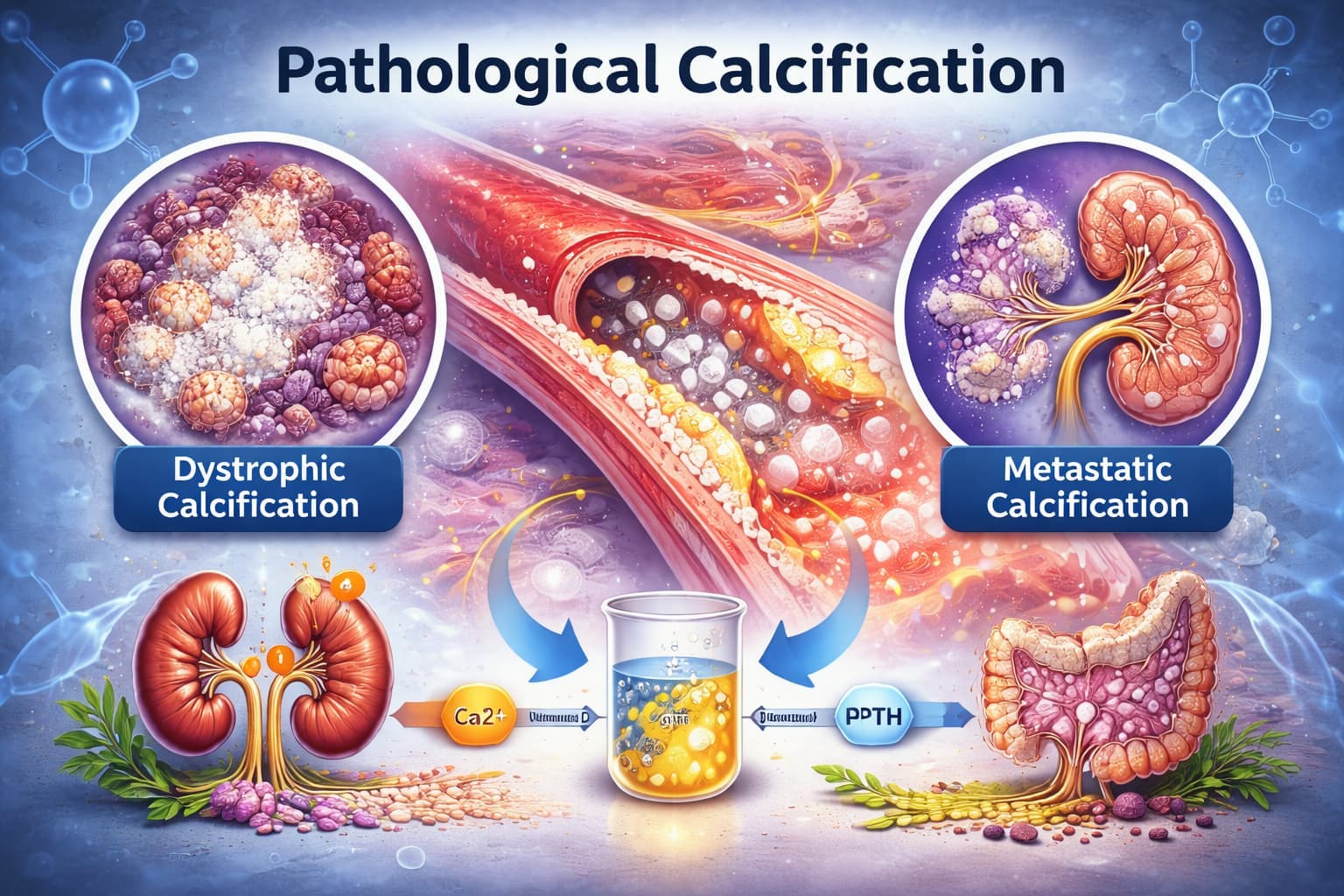
Calcium deposits show up in tissue where they do not belong. When you encounter them on histology or imaging, you are looking at pathological calcification. It is a process that indicates underlying tissue damage, metabolic disruption, or a condition the body has not been able to resolve. It is not a disease in itself, but a morphological marker of disease.
Pathological calcification occurs in two distinctly different contexts: sometimes the tissue is already dead or dying, and sometimes the tissue is entirely normal. But the body's calcium balance has gone haywire.
In pathology practice, distinguishing between these types directly shapes differential diagnosis, informs about systemic disease burden, and in some cases, changes management.
What Is Pathological Calcification?
The definition of pathological calcification is the abnormal deposition of calcium salts, viz., predominantly calcium phosphate and calcium carbonate in soft tissues, organs, or vessels where calcium does not ordinarily accumulate. These deposits may appear as fine granular specks, dense conglomerates, or even bone-like structures.
Pathological calcification is distinct from physiological calcification. The latter occurs during normal bone and tooth mineralisation.
In pathological settings, the process is driven by either local tissue injury or systemic calcium-phosphate imbalance. This distinction forms the foundation of the entire classification.
Types of Calcification in Pathology
Pathology calcification is classically divided into three major categories.
- Dystrophic Calcification.
- Metastatic Calcification.
- Idiopathic (Mönckeberg's) Calcification.
Each type has a distinct biochemistry, anatomical location, and significance.
1. Dystrophic Calcification
Dystrophic calcification pathology is the most common form you will see in routine practice. It occurs in areas of necrosis, degeneration, or dead tissue, even when serum calcium levels are completely normal. The body's calcium does not cause it. It is caused by local tissue changes.
Mechanism
When cells die, especially through coagulative, caseous, or liquefactive necrosis, the plasma membranes lose their ability to regulate intracellular calcium. Calcium floods into the cell.
Simultaneously, phospholipids released from damaged membranes act as nucleation sites for calcium crystal formation. The result is progressive mineralisation of the necrotic focus.
Acidic pH at sites of necrosis initially inhibits deposition. But as alkaline phosphatase activity rises, generated by dying cells and inflammatory macrophages, local phosphate concentrations increase and crystallisation takes hold.
Where You Find It
- Atherosclerotic plaques: Calcium deposits within necrotic lipid cores, contributing to plaque rigidity.
- Calcific aortic stenosis: Progressive calcification of the valve leaflets following decades of mechanical stress and micro-injury. It leads to reduced valve mobility and eventual obstruction.
- Old tuberculosis lesion: Caseous necrotic foci calcify over time; the classic 'Ghon focus' is a prime example.
- Fat necrosis: Post-traumatic or enzymatic fat necrosis, commonly in the breast or pancreatic bed.
- Endometrial calcification pathology: Dystrophic calcification of necrotic endometrial stroma or retained products of conception; these appear as echogenic foci on ultrasound and are a marker of prior endometrial injury or chronic infection.
- Placental calcification pathology: A physiologically accepted variant in term pregnancies, but accelerated calcification in preterm placentas can indicate placental insufficiency, chronic abruption, or vascular compromise.
In dystrophic calcification, serum calcium is always normal. The mineralisation is entirely a local phenomenon.
2. Metastatic Calcification
Despite the name, Metastatic calcification pathology has nothing to do with cancer metastasis. It refers to calcium deposition in normal, previously uninjured tissue, driven by hypercalcaemia or an elevated calcium-phosphate in the blood.
Mechanism
When serum calcium rises above the threshold at which the calcium-phosphate product exceeds approximately 70 mg²/dL², spontaneous precipitation of calcium phosphate occurs in tissues. Tissues that are particularly vulnerable are those with an alkaline local microenvironment.
The main drivers of metastatic calcification include the following.
Primary Hyperparathyroidism: Excess PTH mobilises calcium from bone into blood.
Hypervitaminosis D: Promotes intestinal calcium absorption and renal reabsorption.
Milk-alkali syndrome: Excess calcium and absorbable alkali intake.
Widespread osteolytic metastases: Bone destruction releases calcium into circulation.
Sarcoidosis and granulomatous diseases: Ectopic 1-alpha-hydroxylase activity in macrophages produces excess calcitriol.
Chronic renal failure: Phosphate retention drives calcium-phosphate product elevation.
Preferred Sites
Metastatic calcification gravitates to tissues that excrete acid, creating a relatively alkaline local environment as a by-product. These include:
Renal tubular basement membranes (nephrocalcinosis).
- Pulmonary alveolar walls, presenting as diffuse pulmonary calcification.
- Gastric mucosa, parietal cells secrete HCl, leaving the interstitium alkaline.
- Cornea and conjunctiva (band keratopathy).
- Blood vessel walls, producing 'pipe-stem' calcification.
- Synovial membranes.
Unlike dystrophic calcification, metastatic calcification may be reversible if you identify and treat the underlying cause of hypercalcaemia promptly.
3. Idiopathic (Mönckeberg's) Calcification
Mönckeberg's medial calcific sclerosis represents a third, less well-understood category. In this condition, calcium deposits appear in the tunica media of medium-sized muscular arteries without prior necrosis and without hypercalcaemia. It happens particularly in elderly patients.
Serum calcium is normal. The tissue itself shows no prior injury on standard histology.
The mechanism is not fully elucidated. But current evidence implicates the transformation of vascular smooth muscle cells into osteoblast-like cells under the influence of oxidative stress, ageing, and diabetes-related metabolic signals. These cells then drive mineralisation in an otherwise intact vessel wall.
You usually see it as 'eggshell' or 'railroad track' calcification on imaging, and histologically as rings of calcium in the media without intimal plaque.
It does not obstruct the vessel lumen. But does reduce arterial compliance, contributing to isolated systolic hypertension.
Comparing the Three Types: A Quick Reference
The table below consolidates the distinguishing features of each type of pathological calcification to support faster clinical and histological differentiation.
| Feature | Dystrophic Calcification | Metastatic Calcification | Idiopathic Calcification |
|---|---|---|---|
| Serum Calcium | Normal | Elevated | Normal |
| Underlying Cause | Necrosis / Degeneration | Hypercalcaemia | Unknown |
| Common Sites | Atherosclerotic plaques, old TB lesions, heart valves | Kidneys, lungs, gastric mucosa | Soft tissues, vessel walls |
| Calcium Deposits | In dead/dying tissue | In normal viable tissue | In normal tissue, no clear trigger |
| Reversibility | Usually irreversible | May reverse if cause is treated | Unpredictable |
| Clinical Significance | Marker of prior disease | Indicates systemic metabolic disorder | Often found incidentally |
Pathological Calcification in the Brain
Pathological calcification in the brain deserves separate attention because the CNS is an unusually sensitive environment. When you encounter intracranial calcium deposits, the differential is broad and the clinical implications can be significant.
Pathological calcification sites in the brain include the following.
Basal ganglia: Bilateral basal ganglia calcification is seen in Fahr's disease (primary familial brain calcification), hypoparathyroidism, pseudohypoparathyroidism, and mitochondrial disorders
Choroid plexus: A frequent incidental finding, especially with ageing; generally benign unless asymmetric or extensive
Pineal gland: Physiological in adults. But it should prompt evaluation if pronounced in younger patients
Periventricular and subcortical white matter: Seen in congenital TORCH infections (especially CMV and toxoplasmosis), tuberous sclerosis, and Sturge-Weber syndrome.
Cerebral vessels: Atherosclerotic calcification of intracranial arteries, or calcification associated with prior haemorrhagic infarcts.
Tumour-associated calcification: Oligodendrogliomas, craniopharyngiomas, and meningiomas classically calcify; the pattern and distribution of calcification can be a diagnostic clue
The mechanism in brain calcification is multifactorial. Calcium-phosphate product elevation (as in hypoparathyroidism), mitochondrial dysfunction, and aberrant mineralisation signalling through the SLC20A2 phosphate transporter (mutated in Fahr's disease) all play documented roles. In congenital infections, direct cytopathic effects on periventricular cells create dystrophic foci.
How to Define and Classify in Practice
When you define and classify pathological calcification in a given specimen, a structured approach gets you to the right answer faster. Start with the clinical context. Then apply histology to assess.
- Is the calcification in necrotic, degenerated, or previously normal tissue?
- What is the distribution and pattern of deposits (granular, psammomatous, sheet-like, ring-like)?
- Is there an inflammatory infiltrate or giant cell response?
- Are vascular structures involved, and if so, which layer of the vessel wall?
Von Kossa staining (black deposits) and Alizarin Red S (orange-red deposits) confirm the presence of calcium salts. Electron microscopy reveals hydroxyapatite crystals in advanced cases. Paired with immunohistochemistry and clinical correlation, you can arrive at a precise categorisation that directly supports the treating clinician's decision-making.
A Note on Psammoma Bodies
Psammoma bodies are laminated. They are concentric calcified structures that form through a specific dystrophic mechanism. Successive layers of calcium deposit around a necrotic cell or cellular fragment.
They are not a separate type of pathological calcification, but rather a morphological variant of dystrophic calcification with strong diagnostic associations.
- Papillary thyroid carcinoma
- Papillary serous ovarian carcinoma
- Meningioma
- Papillary renal cell carcinoma
When you spot psammoma bodies, let the clinical and histological context guide the diagnosis. They are a clue, not a diagnosis in themselves.
Flabs LIS: AI-Powered Pathology Lab Software
Managing complex diagnostic findings like pathological calcification requires more than good clinical judgment. You need a lab system built for precision. Flabs LIS brings AI-powered intelligence directly into your reporting process.
AI Voice Reporting: Dictate your calcification findings naturally; AI converts them into structured, accurate diagnostic reports instantly.
AI Interpretation: Auto-generate contextual interpretations for calcium-related findings, reducing manual effort and human error.
AI Smart Report: Deliver polished, standardised reports for every case, from routine calcification screens to complex metastatic workups.
AI Flagger: Automatically flag abnormal calcium markers and critical results before reports leave your lab.
TAT Management: Track every calcification workup in real time and ensure on-schedule report delivery every single time.
Quality Control (QC): Automated checks ensure every calcification report meets NABL and industry standards before delivery.
Dynamic QR Code & Barcode: QR-coded, tamper-proof reports with instant soft copy access and barcode-based sample tracking.
Bulk Actions: Bulk registration, report download, approvals, invoicing, and WhatsApp/SMS/Email sharing.
Lab Finance Management: Automate invoicing, payments, and financial records with complete accuracy.
With AI-powered features built for scalable pathology labs, FLABS can transform your lab operations, adhering to certified medical standards.
Start Your 5-Day Free Trial Now!
Also read - Types of Crystals in Urine and Their Significance
Get Started at ₹1!
Try Flabs for a full month for just ₹1.
Follow us on