Pathologists often have to pull a urine sample under the microscope. Something that is centrifuged, stained and ready. As they scan the field, they sometimes spot crystals.
Some are common, some are not. Some are clinically silent, others indicate a metabolic disorder. Your interpretation can shape the next step for the patient, the clinician, and the diagnosis. So, let’s talk about the types of crystals in urine you encounter daily, what they mean, and how to distinguish one from another without second-guessing.
What are Urine Crystals and Why Do they Matter?
Urine is a complex solution. It carries salts, minerals, and organic compounds. When urine pH, temperature, or concentration shifts, those dissolved substances can precipitate. That’s when you see crystals. And under the microscope, they reveal clues.
Now, not every crystal is pathological. Some are simply dietary. Some indicate a sample sat too long at room temperature. But others point to liver disease, inherited disorders, or acute kidney injury. Knowing the types of crystals in urine microscopy means knowing the difference between artefacts and emergencies.
Common Types of Crystals in Urine Microscopy
| Crystal Type | Shape/Appearance | Urine pH | Clinical Association |
|---|---|---|---|
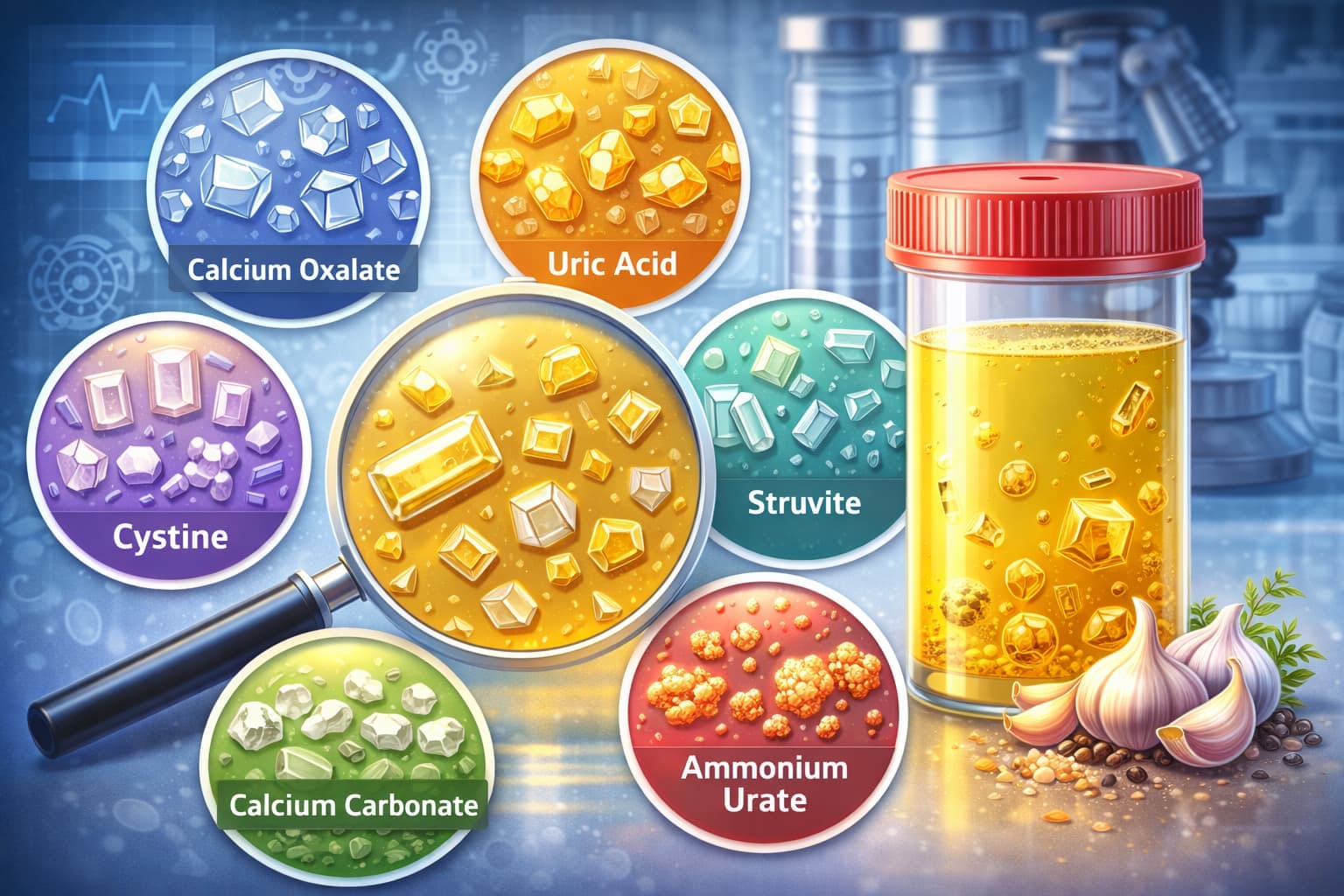
| Urate crystals | Variable: barrels, rosettes, rhomboids, needles, hexagonal plates; amber coloured | Usually acidic | Seen occasionally in normal urine; common in urate nephrolithiasis |
| Cystine crystals | Colourless hexagonal plates (benzene-ring appearance) | Acidic (<6) | Cystinuria (genetic defect of cystine transport) |
| Triple phosphate (Struvite) | Coffin-lid shape | Alkaline (>7) | UTI with urea-splitting bacteria (e.g., Proteus); struvite stones |
| Calcium oxalate | Dihydrate: envelope shape; Monohydrate: dumbbell, rod, oval | Usually acidic | Nephrolithiasis; ethylene glycol poisoning |
| Calcium carbonate | Large spheroids with radial striations | Variable | Mostly in herbivores; rare in humans |
| Amorphous crystals | Fine granular aggregates, no definite shape | Urates → acidic; Phosphates → alkaline | Usually no clinical significance; may mimic bacteria |
| Ammonium biurate | Brown spherical “thorn-apple” crystals | Neutral to alkaline | Seen in liver disease/portal shunts, urate stones |
| Bilirubin crystals | Yellow needle-like or granular crystals | Acidic | Seen in bilirubinuria; liver or biliary disease |
| Drug-induced crystals | Sulfadiazine: sheaves; Indinavir: needle clusters | Variable | Drug therapy; may cause crystal-induced renal failure |
Uric Acid Crystals in Urine
These are among the most common you’ll see. They’re pleomorphic, i.e., they can look like diamonds, rosettes, or even barrels. Colour? Yellow to reddish-brown. They are found in acidic urine, usually with a pH below 5.5.
If you spot uric acid crystals in urine, consider the patient’s hydration status first. Dehydration concentrates the urine, and uric acid precipitates. But recurrent findings? Think gout, tumour lysis syndrome, or high-purine diets. In pathology labs, these crystals are routine but worth noting, especially if accompanied by hematuria or flank pain.
Calcium Oxalate Crystals
Now, this is where morphology gets interesting. You have two main players.
First, calcium oxalate dihydrate crystals in urine. These appear as octahedra, like envelopes. Very distinct. You see them in acidic to neutral urine. They’re often dietary, linked to spinach, rhubarb, or vitamin C.
Then you have the monohydrate form. These are dumbbell or oval-shaped. Harder to spot, but clinically louder. The types of calcium oxalate crystals in urine matter because the monohydrate form is associated with ethylene glycol poisoning. Yes, antifreeze. So if you see those, don’t ignore them.
And broadly, oxalate crystals in urine can signal enteric hyperoxaluria or Crohn’s disease. Always correlate with clinical history.
Amorphous Crystals in Urine
Amorphous. It means without form. And that’s exactly what these look like: shapeless granules. But colour gives them away.
Amorphous phosphate crystals in urine appear as colourless or white granules. They form in alkaline urine. Clinically, they’re benign. Usually just a sign of a vegetarian meal or a urine sample that sat too long.
Amorphous crystals in the urine of the urate variety? Those are yellow or brick-dust coloured. Found in acidic urine. Again, mostly insignificant unless persistent, which might indicate chronic kidney disease.
You’ll see these daily. They’re background noise. But if you’re scanning types of crystals in urine microscopy, you still need to document them. Because pattern matters.
Triple Phosphate Crystals
These are coffin-lid shaped. Literally. Rectangular prisms with oblique ends. Found in alkaline urine. They’re composed of magnesium, ammonium, and phosphate.
Seen them? Usually associated with urinary tract infections caused by urea-splitting bacteria like Proteus. If you report these, the clinician should investigate for infection or struvite stones.
Cystine Crystals in Urine
Now we’re talking about something rare. Cystine crystals in urine look like benzene rings, i.e., hexagonal, colourless, and flat. They form in acidic urine.
If you see these, stop. This isn’t dietary. It’s cystinuria, an inherited disorder. Patients can’t reabsorb cystine, so it accumulates and forms stones. Early detection saves kidneys. So when you spot these, flag them. Immediately.
Cholesterol Crystals
These look like notched, rectangular plates. Sometimes stacked. You’ll see them in alkaline or neutral urine. Cholesterol crystals are rare. But when present, think of nephrotic syndrome or chyluria. They indicate lipiduria.
Not something you see every day. But knowing the types of crystals in urine includes these oddities keeps your differential sharp.
Crystals That Mimic Each Other
Here’s where experience kicks in. Sulfonamide crystals can look like dark-brown spheroids. They’re drug-induced. Rare now, but still possible. Leucine crystals? Spherical with radial striations. Seen in liver disease. Tyrosine crystals? Fine needles. Also liver-related.
You can’t rely on morphology alone. pH, birefringence under polarised light, and patient history complete the picture. That is why pathology labs invest in quality lab equipment, good optics, polarised filters, and consistent reagents.
Clinical Significance: When Crystals Matter
Crystals can be innocent bystanders. But they can also be the first sign of systemic disease.
- Uric acid crystals in serial samples? Gout or myeloproliferative disorders.
- Cystine crystals? Refer to nephrology.
- Calcium oxalate monohydrate with high anion gap metabolic acidosis? Consider ethylene glycol ingestion. It is time-sensitive and life-threatening.
- Amorphous phosphate crystals in urine with recurrent UTIs? Might indicate chronic infection.
Microscopy Best Practices
You already know this, but let’s reinforce. Fresh samples are non-negotiable. Old samples form crystals artificially. Also, note the pH. Always. It guides identification.
If you’re unsure, use polarised light. Cholesterol and cystine are birefringent. Uric acid can be, too. And if you’re still uncertain, correlate with clinical data. The types of crystals in urine don’t exist in a vacuum. They live in the context of the patient.
The Bigger Picture
Knowing about different types of crystals in urine, you’re interpreting a complex chemical story. Every sample tells you something, either about hydration, metabolism, medication, or disease. While they're clues, your expertise turns those clues into answers.
So next time you are at the scope, take a moment. Look closely. That amorphous clump might be nothing. Or it might be the first sign of something bigger. Either way, you’ll know. Because you’ve seen them all. You’ve read the patterns. And you’ve got the tools to document, report, and act.
That’s the difference between a lab that processes samples and a lab that drives diagnosis.
The Role of Lab Automation
Identifying the types of crystals in urine microscopy requires careful observation and clear documentation.
In busy diagnostic laboratories, translating these microscopic findings into structured, clinically meaningful reports can take significant time and coordination between technicians and pathologists. As pathology labs increasingly focus on automation, a reliable lab software that assists you in the process.
Platforms like FLABS AI-Powered Pathology Lab Software help streamline the whole process. It supports structured reporting, AI-assisted interpretations, automated report delivery, and real-time tracking of TAT .
This allows laboratories to focus on accurate diagnosis. It also ensures reports reach clinicians and patients quickly and securely.
Trusted by 2000+ diagnostic labs, FLABS is built to simplify everyday lab operations.
Explore how FLABS supports efficient and accurate lab reporting workflows.
Start Your 5-Day Free Trial Today
Also check - Urine Cytology Test: How It Helps Detect Abnormal Cells
Get Started at ₹1!
Try Flabs for a full month for just ₹1.
Follow us on