Flabs
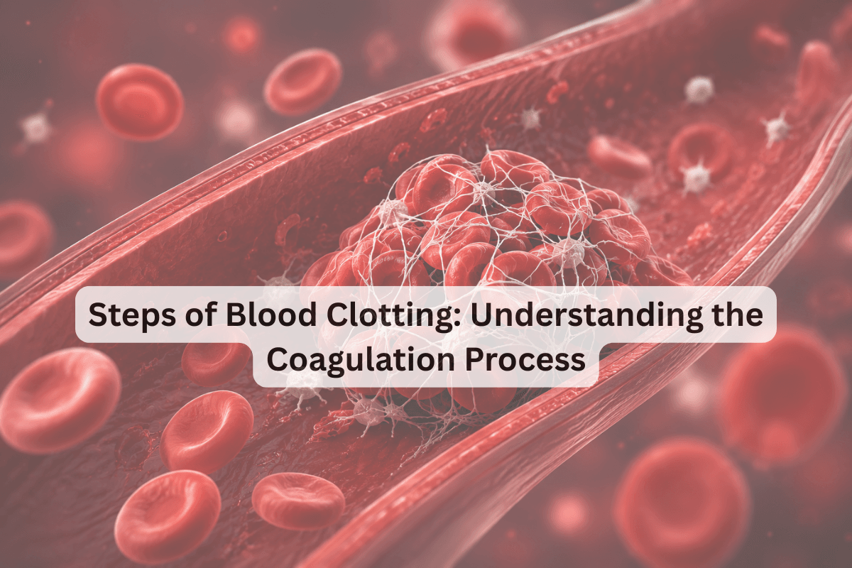
Steps of Blood Clotting: Understanding the Coagulation Process
When a blood vessel breaks, your body launches a precisely sequenced biological response to stop bleeding. That response is the coagulation process. It is one of the most intricate, multi-protein cascades in human physiology.
As a pathologist or lab professional, you already run coagulation tests daily. But having a sharp, refreshed mental model of what you're actually measuring, and why each step matters clinically, makes you a better interpreter of results. Let's understand the steps of blood clotting from first principles.
What Are the Major Steps of Blood Clotting?
The steps of blood clotting occur in four sequential, overlapping phases: vascular spasm, primary haemostasis (platelet plug formation), secondary haemostasis (the coagulation cascade), and fibrinolysis.
Each phase depends on the successful completion of the one before it. A failure at any point shows up in your coagulation tests.
Phase 1: Vascular Spasm
The moment a vessel sustains injury, the smooth muscle in the vessel wall contracts immediately. This vascular spasm reduces blood flow to the injured area within seconds. It buys the body time to mount a more structured haemostatic response.
The spasm is triggered by pain signals from damaged tissue, local myogenic reflexes, and chemical mediators released from platelets and the injured endothelium, particularly endothelin and thromboxane A₂.
At this step of blood clotting, no clotting factor is activated yet. It is purely a mechanical, reflex-driven response.
Phase 2: Primary Haemostasis: Platelet Plug Formation
Once the vessel wall is breached, subendothelial collagen is exposed. This sets off three platelet responses in rapid succession.
Platelet Adhesion
von Willebrand factor (vWF) bridges exposed collagen and the glycoprotein Ib (GPIb) receptor on the platelet surface. This tethers platelets to the injury site despite blood flow shear forces. A deficiency in vWF disrupts this step entirely, as in von Willebrand disease.
Platelet Activation
Adherent platelets activate. They release the contents of their alpha and dense granules, viz., ADP, thromboxane A₂, serotonin, and coagulation factors. ADP and thromboxane A₂ act in a positive feedback loop, recruiting and activating more platelets to the site.
Platelet Aggregation
Activated platelets express GPIIb/IIIa receptors on their surface. Fibrinogen cross-links these receptors between adjacent platelets, forming a loose platelet plug. This is primary haemostasis. It stops minor bleeding but remains unstable without the reinforcement of secondary haemostasis.
Clinical note: Platelet function analysers and bleeding time tests assess this phase. Aspirin and clopidogrel both target platelet activation at this level.
Phase 3: Secondary Haemostasis: The Coagulation Cascade
Secondary haemostasis stabilises the platelet plug by generating a fibrin mesh through a series of enzymatic reactions. It is known as the coagulation cascade.
You can describe the process of blood clotting in human beings through two converging arms: the extrinsic pathway and the intrinsic pathway, both of which feed into the common pathway.
The Extrinsic Pathway (Tissue Factor Pathway)
Tissue factor (TF), expressed on subendothelial cells and monocytes, is exposed at injury. TF binds circulating Factor VIIa, forming the TF-VIIa complex. This complex activates Factor X directly and also activates Factor IX, linking the two pathways. The extrinsic pathway is fast and is measured by the Prothrombin Time (PT/INR) on your coagulometer.
The Intrinsic Pathway (Contact Activation Pathway)
Exposed collagen and negatively charged surfaces activate Factor XII (Hageman factor), which cascades through Factor XI → Factor IX → Factor VIII. The intrinsic pathway among the steps of blood clotting is slower but amplifies the coagulation signal significantly. It is measured by the Activated Partial Thromboplastin Time (aPTT).
The Common Pathway
Both pathways converge at Factor X activation. Factor Xa, together with Factor Va (the prothrombinase complex), converts prothrombin (Factor II) to thrombin. Thrombin then converts soluble fibrinogen to insoluble fibrin monomers. Factor XIII, activated by thrombin, cross-links fibrin monomers into a stable, resilient clot.
| Pathway | Factors Involved | Lab Test | Measured By |
|---|---|---|---|
| Extrinsic | TF, Factor VII, Factor X | PT/INR | Coagulometer |
| Intrinsic | Factors XII, XI, IX, VIII | aPTT | Coagulometer |
| Common | Factors X, V, II (Prothrombin), I (Fibrinogen) | PT, aPTT, TT | Coagulometer |
| Primary Haemostasis | Platelets, vWF, GPIb, GPIIb/IIIa | Platelet count, PFA-100, BT | Analyser/Manual |
A well-calibrated coagulometer in your lab is the single most important piece of lab equipment for quantifying secondary haemostasis.
Instrument calibration, reagent lot consistency, and sample pre-analytics all directly affect PT and aPTT accuracy.
Pathology labs in India dealing with high-volume haematology workloads need tight quality control (QC) protocols at every run. Especially when managing anticoagulation monitoring.
Phase 4: Fibrinolysis: Clot Resolution
Once tissue repair begins, the clot must be dissolved to restore normal blood flow. Plasminogen, embedded in the fibrin clot, is activated to plasmin by tissue plasminogen activator (tPA) released from endothelial cells. Plasmin degrades the fibrin mesh into fibrin degradation products (FDPs) and D-dimers.
D-dimer measurement is a direct read on fibrinolytic activity. Elevated D-dimers indicate active clot formation and breakdown. It is a major marker in DIC, DVT, and pulmonary embolism workups.
Natural anticoagulants (antithrombin III, Protein C, and Protein S) simultaneously inhibit clotting factors to prevent the cascade from running unchecked.
Where the Steps of Blood Clotting Break Down
Every clotting disorder maps to a specific step in this sequence. Here's how defects present in your coagulation tests:
| Disorder | Affected Step | Abnormal Test Results |
|---|---|---|
| Haemophilia A | Factor VIII deficiency (Intrinsic pathway) | Prolonged aPTT, Normal PT |
| Haemophilia B | Factor IX deficiency (Intrinsic pathway) | Prolonged aPTT, Normal PT |
| Vitamin K Deficiency | Factors II, VII, IX, X are impaired | Prolonged PT and aPTT |
| DIC | Systemic activation + fibrinolysis | ↑ PT, ↑ aPTT, ↑ D-dimer, ↓ Fibrinogen, ↓ Platelets |
| von Willebrand Disease | Platelet adhesion (Primary haemostasis) | Prolonged BT/PFA, ± aPTT |
| Liver Disease | Reduced factor synthesis | Prolonged PT (early), then prolonged aPTT |
Recognising which pathway is affected based on which coagulation tests are abnormal is the core of haemostasis interpretation. Hence, the steps of blood clotting are the diagnostic map you use every single day.
Medications for Blood Clotting
| Category | Mechanism of Action | Medications |
|---|---|---|
| Antiplatelet Drugs | Prevent platelets from sticking together and forming clots | Aspirin, Clopidogrel, Dipyridamole, Prasugrel, Ticagrelor, Ticlopidine |
| Anticoagulants (Blood Thinners) | Inhibit clotting factors or proteins involved in clot formation | Apixaban, Dabigatran, Edoxaban, Heparin, Rivaroxaban, Warfarin |
| Thrombolytics (Clot-Dissolving Drugs) | Activate proteins that break down fibrin strands to dissolve clots | Alteplase, Streptokinase, Tenecteplase |
Accurate Coagulation Reporting
The steps of blood clotting span seconds to minutes in the body. But errors in lab reporting can delay clinical decisions for hours. For NABL labs, coagulation test reporting demands both analytical precision and speed.
Pre-analytical errors are the leading cause of spurious PT and aPTT results, viz., haemolysis, incorrect citrate ratio, and delayed processing. Your lab's quality control systems must flag these before reports go out.
When labs run high volumes of coagulation tests, manual oversight at every step becomes impractical. This is where intelligent lab information systems make a measurable difference in turnaround time and result accuracy.
FLABS LIS for Modern Pathology Labs
High-volume coagulation testing demands more than accuracy. It requires speed, standardisation, and zero tolerance for error. FLABS LIS is built specifically for modern pathology labs that need to scale without compromising quality.
From automated QC checks on PT/INR and aPTT runs to intelligent flagging of critical results, FLABS ensures every report that leaves your lab is both reliable and audit-ready. Real-time turnaround time (TAT) tracking, instrument integration, and structured data operations help you maintain NABL compliance without manual bottlenecks.
Whether you're managing anticoagulation monitoring, handling bulk test loads, or reducing pre-analytical errors, FLABS gives you operational control and clinical confidence at scale.
Start your 5-day free trial today and see how FLABS LIS can optimise your lab operations from sample intake to final report delivery.